
Graves' disease - Case 12. (ultrasonographic picture 5)
|
|

|
|
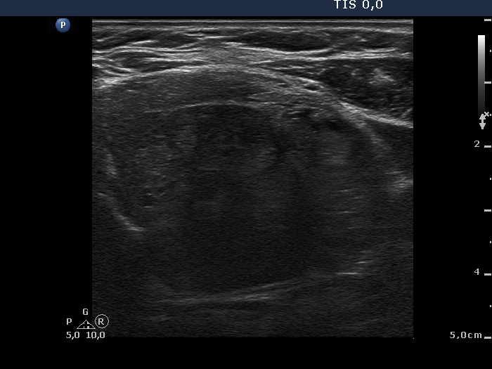
Left lobe, horizontal scan. The pattern is basically similar to that demonstrated in the right lobe. However, there are several less hypoechogenic discrete lesions within the central hypoechogenic area. These may presentations of hyperplastic nodules.