
Parathyroid lesions - Case 8.
Follow-up 3 years later (ultrasonographic picture 4)

|

|

|
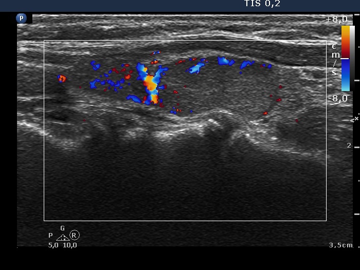
Right lobe, longitudinal scan, color Doppler mode. The larger mass presents a combined type 2 and type 3 pattern.


