
Parathyroid lesions - Case 2.
A patient with a multinodular goiter (ultrasonographic picture 12)
|
|

|
|
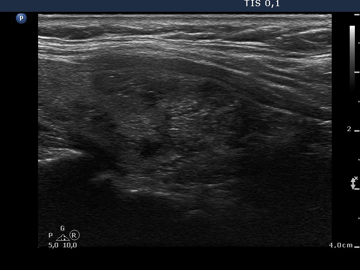
Left lobe, longitudinal scan. Multiple nodules are demonstrated. The synchronous presence of hyperechogenic lines and granules proves that these figures correspond to connective tissue.