
|
|
Subacute granulomatous de Quervain's thyroiditis - Case 42.
|
|

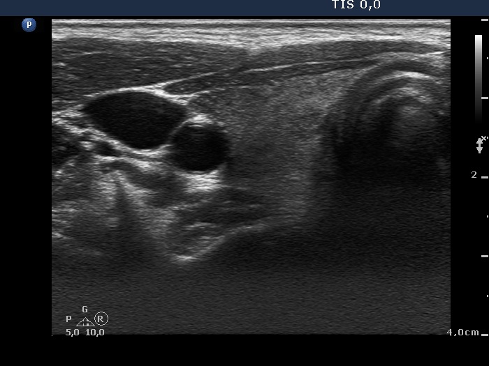


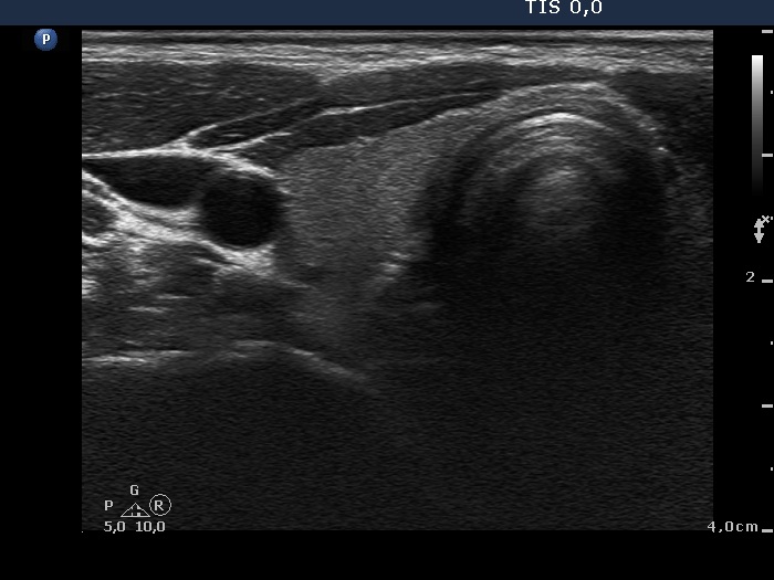
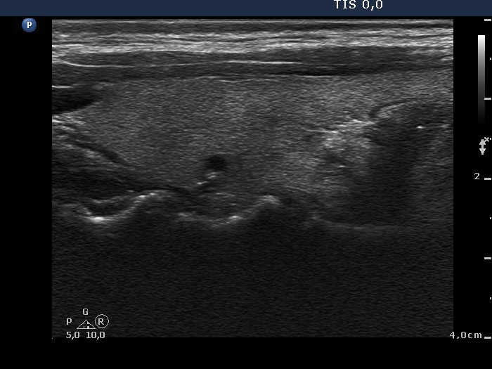
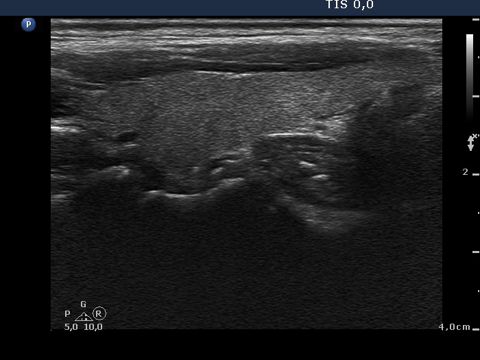
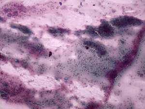
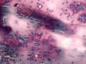
First examination (1st and 4th rows of images):
Clinical presentation: a 34-year-old woman was referred for an evaluation of neck complaints. She suffered from neck pain and fever for two months lasting for 3 weeks. Thereafter complaints suggesting hyperthyroidism have developed. The neck complaints have decreased. The body temperature has normalized.
Palpation: the right thyroid was tender and hard on palpation.
Functional state: hyperthyroidism with TSH 0.01 mIU/L and FT4 27.2 pM/L, CRP 12.8 mg/L (normal value 0-4.8), TSAb 0 U/L.
Ultrasonography: the thyroids were echonormal and presented hypoechogenic areas with ill-defined borders. The vascularization was average.
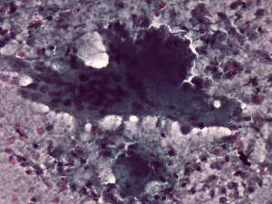
Aspiration cytology: was performed from right thyroid and resulted in subacute, granulomatous, de Quervain's thyroiditis.
Non-steroid anti inflammatory drug was administered.
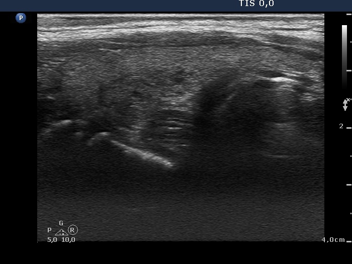
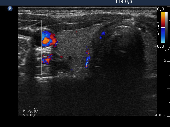
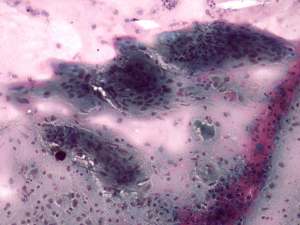
Follow-up examination 3 months later (2nd row of images):
Clinical presentation: the complaints of the patient ceased.
Palpation: the thyroid was not tender, both lobes were firm.
Functional state: hypothyroidism with TSH 2.98 mIU/L and FT4 7.56 pM/L. CRP 1.9 mg/L.
Ultrasonography: the proportion of hypoechogenic areas has decreased. The vascularization became normal.
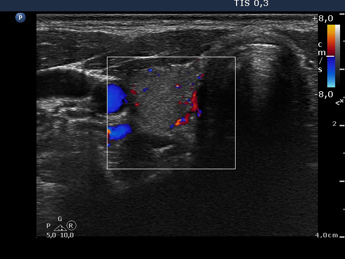
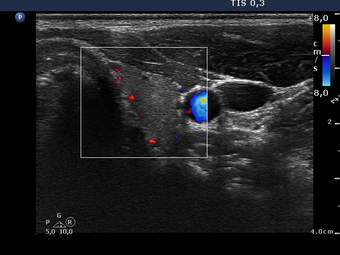
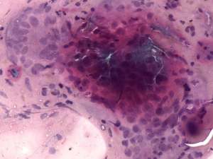
Follow-up examination 11 months later (3rd row of images):
Clinical presentation: the patient had no complaints.
Palpation: no abnormality.
Functional state: subclinical hyperthyroidism with TSH 1.09 mIU/L, and FT4 17.1 pM/L.
Ultrasonography: the thyroid was echonormal with several small minimally hypoechogenic areas. The vascularization was less than the average.
.