
The operated thyroid - Case 20. (ultrasonographic picture 3)
A previously operated patient with a newly developed Graves' disease

|

|

|
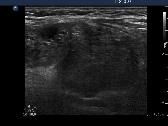
Lower part of the right lobe, longitudinal scan. The discrete hypoechogenic areas mimic a multinodular goiter. But in contrast with a nodule in a pathological sense these lesions have irregular borders.


