
|
|
Graves' disease - Case 14.
|
|


Clinical data: A 31-year-old woman was referred for evaluation of a suspicious nodule. She was treated for Graves' hyperthyroidism for 15 months. At the time of the discontinuation of the thyrostatic therapy, an ultrasound examination was performed, and a hypoechogenic nodule with microcalcifications was described.
Palpation: no abnormality.
Results of blood tests: euthyroidism (TSH 2.98 mIU/L, FT4 11.0 pM/L).
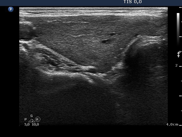
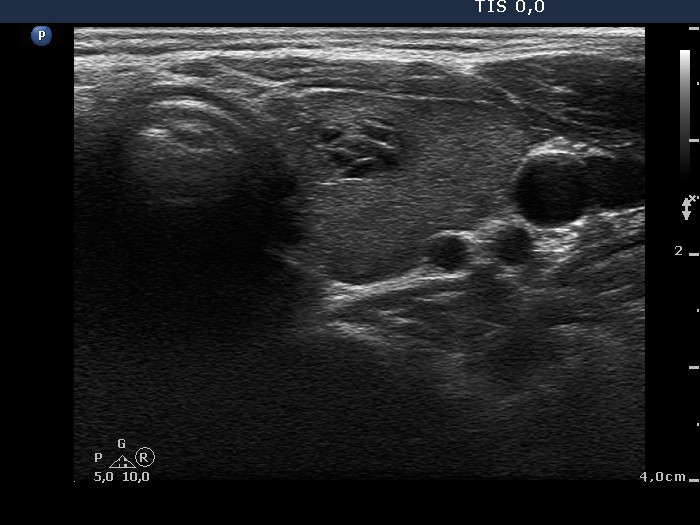
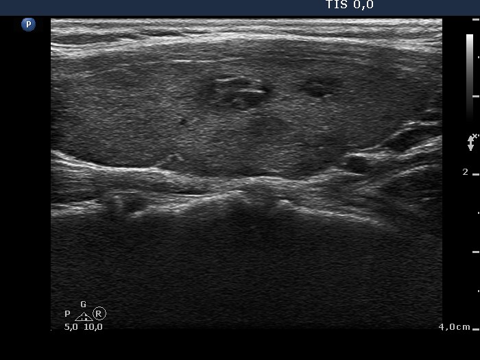
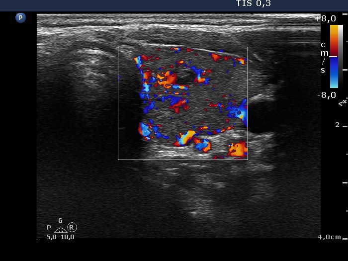
Ultrasonography: The thyroid was minimally-moderately hypoechogenic with several insignificant discrete areas. There were two hypoechogenic small lesions close to each other within the left lobe. The larger one displayed hyperechogenic lines and figures in the back wall of cystic areas; these are optical artifacts caused by posterior back wall enhancement.
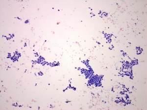
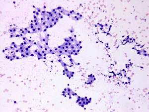
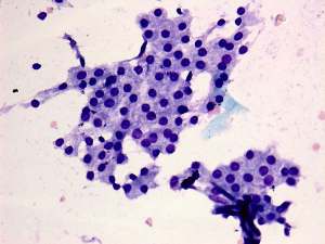
Cytology was performed from the hypoechogenic lesion in the left lobe and resulted in benign lesion with hormonal influences.
Comment. The optical artifacts caused by posterior back wall enhancement should not be interpreted as microcalcifications.