
|
|
Chronic lymphocytic thyroiditis - Case 12.
|
|


Clinical presentation: a 33-year old woman was referred for an evaluation of a goiter evolved over 6 months.
Functional state: hypothyroidism (TSH 21.8 mIU/L, FT4 8.32 pM/L).
Palpation: both thyroids were enlarged and very firm. No nodule was palpable.
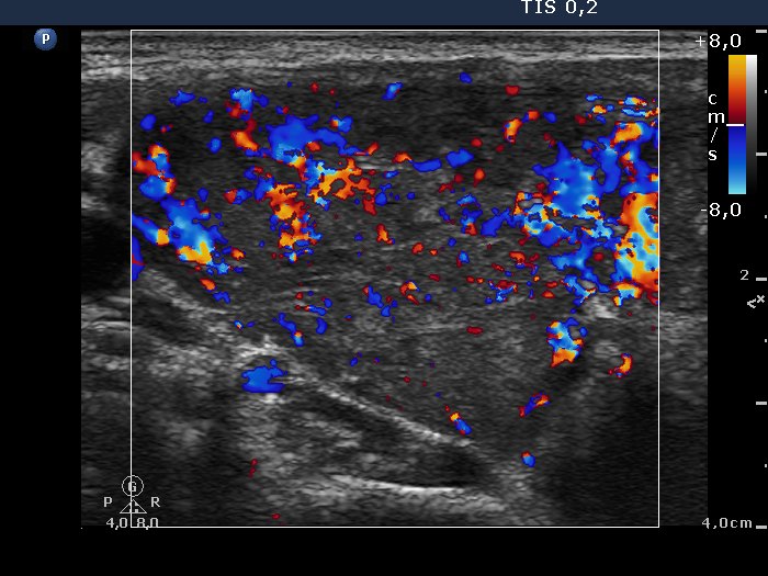
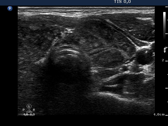
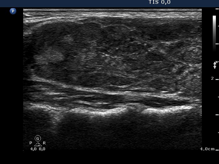
Ultrasonography: the thyroids were hypoechogenic and contained numerous discrete echonormal lesions divided by fibrous tissue. The vascularization was increased.
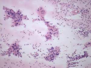
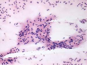
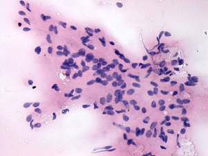
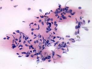
Cytology: was performed and resulted in thyroiditis. There were scattered number of lymphocytes and multinucleated giant cells were also found.
Anti-TPO was determined and was above 900 U/L.
Final combined diagnosis: primary hypothyroidism caused by autoimmune thyroiditis.
Comments:
-
The sonographic pattern corresponds to the so-called micronodular (or pseudonodular) form of lymphocytic thyroiditis.
-
The presence of multinucleated giant cells composed of epitheloid cells resembles that observed in de Quervain's thyroiditis. This type of multinucleated cells occurs rarely in autoimmune thyroiditis.