
|
|
Oxyphilic adenoma - Case 12.
|
|


Clinical presentation: a 29-year-old woman requested a re-evaluation of her nodule known for 3 years. On first occasion and 1 year later she rejected FNAC.
Palpation: a firm nodule in the left lobe.
Functional state: euthyroidism with TSH 1.70 mIU/L.
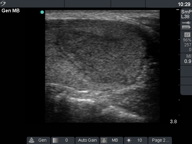
Ultrasonography: thee basic echo structure of the thyroid was normal. There was a hypoechogenic inhomogeneous nodule in the left lobe. The nodule increased in size from 12x10x16 mm to 17x12x19 mm one year after (first row of US images) and to 30x16x29 mm 3 years after the first investigation (second row of US images), i.e. from 1.0 mL to 7.3 mL. Both the perinodular and the intranodular blood flow was increased, the latter irregularly increased.
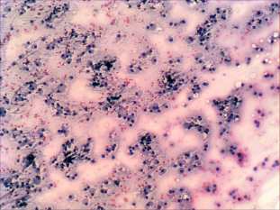
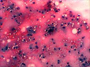
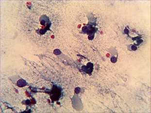
Cytological picture: there is no colloid in the background. Thyrocytes are dispersed. Around 90% of follicular cells exhibit oxyphilic metaplasia. The nuclei do not contain prominent nucleoli .
Cytological diagnosis: Hürthle-cell tumor.
Histopathology disclosed Hürthle-cell adenoma.
Comments:
-
It is a rare situation that we can observe a thyroid tumor over years. (One has practice with those patients whose first FNAC was benign, and a tumor was diagnosed only on a second occasion.) Such an extreme increase in volume of a non-cystic nodule like in this patient is very rarely found and is itself suspicious for malignancy.
-
The pleomorphism has only little relevance in the case of oxyphilic cells arising from either Hashimoto`s thyroiditis or Hürthle-cell tumor.