
|
|
The role of complex diagnosis - follow-up of follicular lesions - Case 1.
|
|


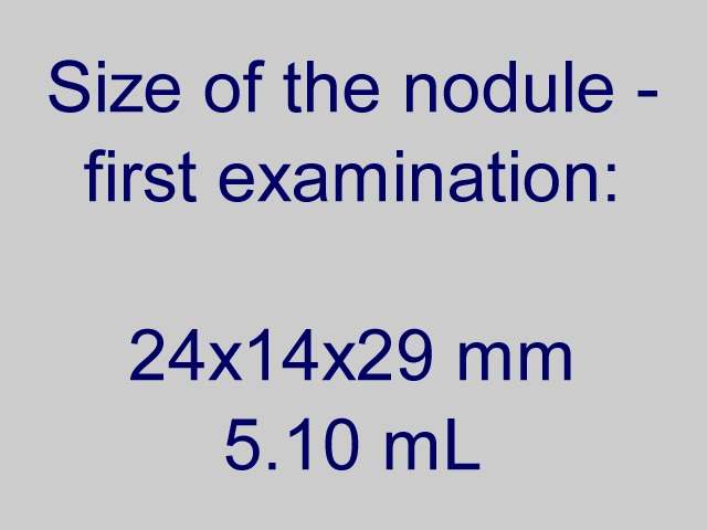
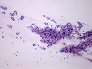
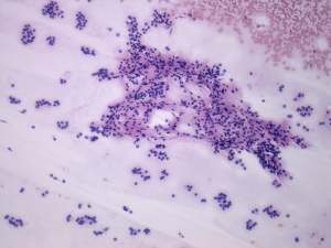

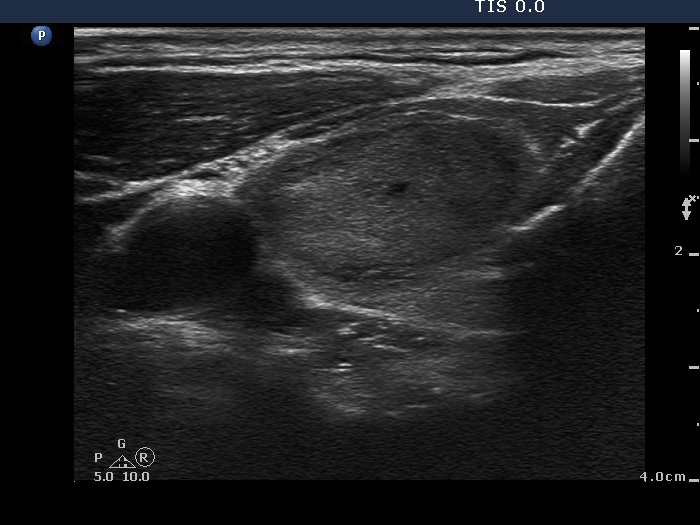
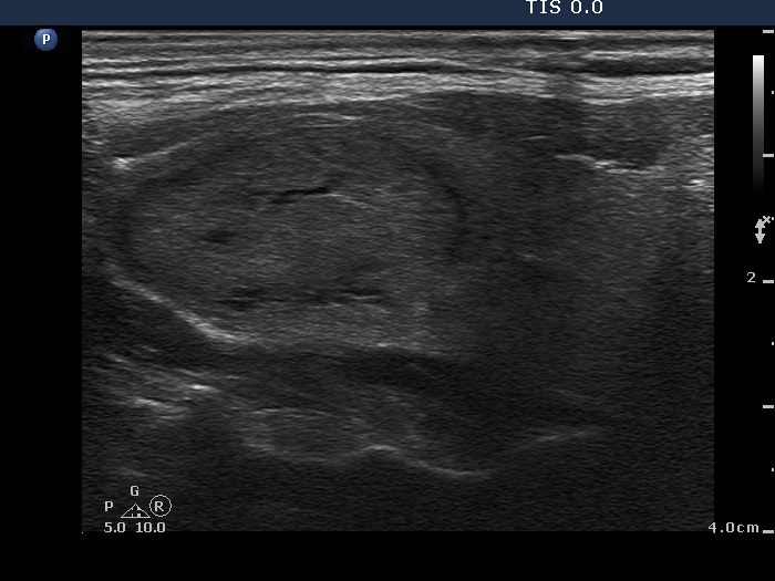
First examination (1st and 2nd rows of images)
Clinical presentation: a 66-year-old man was referred for aspiration cytology. He noticed a lump in the right side of the neck for three weeks. Scintigraphy disclosed a "cold" nodule.
Palpation: a moderately firm nodule in the right lobe.
Functional state: euthyroidism with TSH-level 1.19 mIU/L.
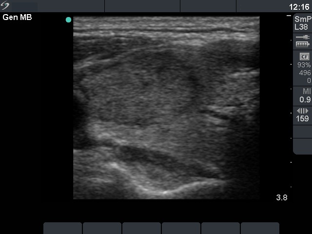
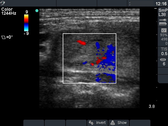
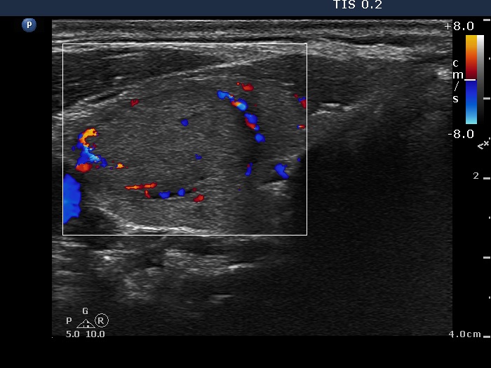
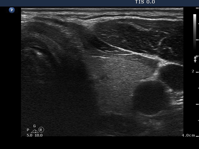
Ultrasonography: the thyroid was echonormal. There was a moderately hypoechogenic nodule in the ventrolateral part of the right lobe. The nodule displayed halo sign and a combined type 2 and type 3 vascular pattern.
Cytology was performed and resulted in follicular tumor without significant atypia.
A combined clinical-ultrasound-cytological diagnosis was follicular tumor with less than 2% risk of carcinoma.
The patient decided to undergo follow-up instead of immediate surgery.
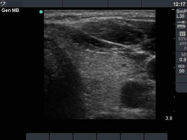
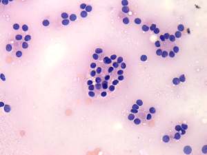
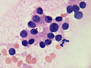
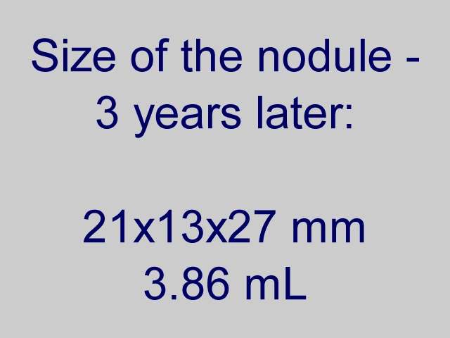
Second examination 3 years later (3rd row of images)
Summary of follow-up: the patient underwent yearly ultrasound examination. The nodule was unchanged, he had no complaints.
Functional state: euthyroidism with TSH-level 2.69 mIU/L.
Ultrasonography: the ultrasound presentation of the thyroid was unchanged.
The nodule has decreased significantly in size over 3 years, the volume of the nodule was 5.10 and 3.86 mL, at the first examination and at the 4-year follow up, respectively.Suggestion: to continue the follow-up with ultrasound and TSH determinations every year.