
|
|
The role of complex diagnosis - follow-up of follicular lesions - Case 2.
|
|


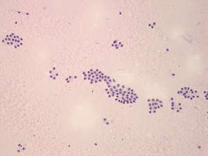

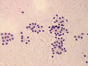
First examination (1st and 2nd rows of images)
Clinical presentation: a 29-year-old woman was referred for evaluation of infertility.
Palpation: a moderately firm nodule in the left lobe.
Functional state: euthyroidism with TSH-level 1.56 mIU/L.
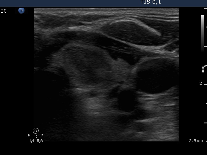
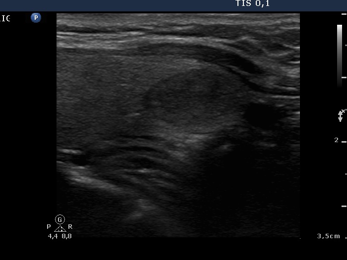
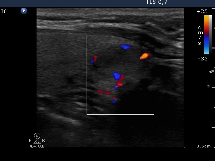
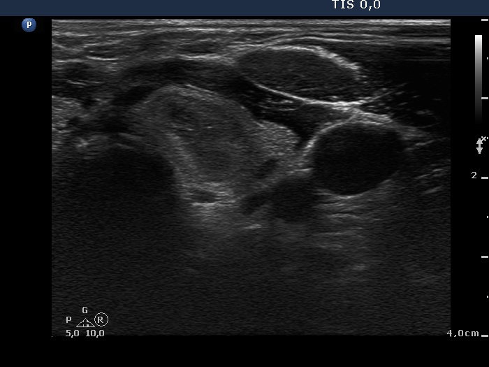
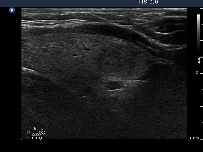
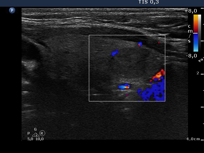
Ultrasonography. The thyroid was echonormal. There was a nodule in the lower pole of the left lobe. The nodule contained echonormal and moderately hypoechogenic areas and presented a halo sign. There were signs of a type 2 vascular pattern.
Cytology was performed and resulted in follicular tumor without significant atypia.
A combined clinical-ultrasound-cytological diagnosis was follicular tumor with less than 1% risk of carcinoma.
The patient decided to undergo follow-up instead of immediate surgery.
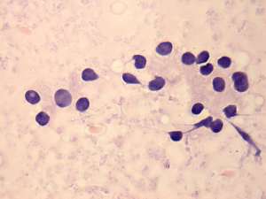
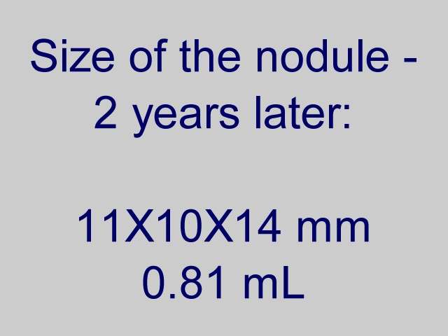
Second examination 2 years later (3rd row of images)
Summary of follow-up: the patient underwent yearly ultrasound examination. The nodule was unchanged, he had no complaints.
Functional state: euthyroidism with TSH-level 1.49 mIU/L.
Ultrasonography: the ultrasound presentation of the thyroid was unchanged.
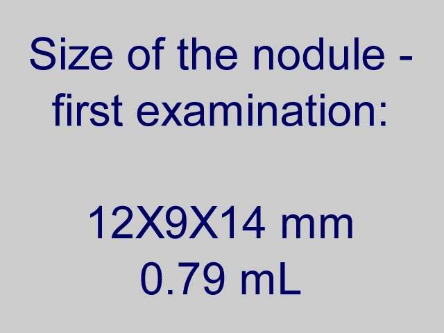
The volume of the nodule was 0.79 and 0.81 mL, at the first examination and at the 2-year follow up, respectively.Suggestion: to continue the follow-up with ultrasound and TSH determinations every year.