
Graves' disease - Case 29.
One year after the first visit (ultrasonographic picture 2)

|

|
 |
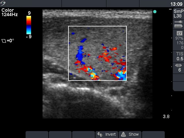
Right lobe, horizontal scan, color Doppler mode. The vascularization is a little bit greater than the average.


