
Differentiation of discrete lesions - case p035 doi: 10.24390/thyrocaseconp035ln.12
Present examination (ultrasonographic picture 6)
|
|

|
|
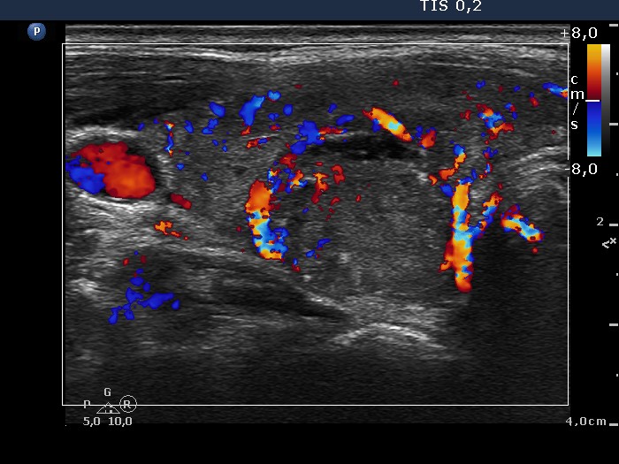
Lower part of the right lobe, horizontal scan, color Doppler mode. The intralesional vascularization is less pronounced compared with the non-lesional part of the lobe.