
|
|
100 consecutive patients with papillary carcinoma - Case 36.
|

|


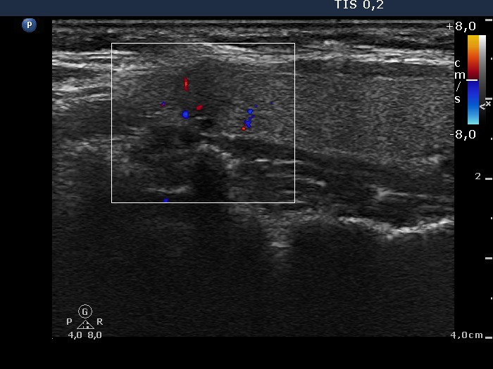
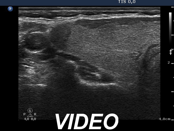


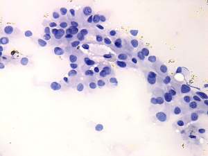
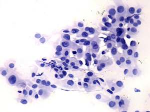
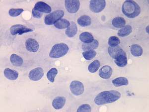
Clinical data: A 40-year-old woman requested evaluation of complaints suggesting hypothyroidism. She was diagnosed with goiter 10 years ago, when scintigraphy disclosed no nodule. No thyroid investigation was made in the previous years.
Palpation: no abnormality.
Hormonal evaluation indicated euthyroidism with TSH 1.96 mIU/L.
Ultrasonography: The thyroid was echonormal. A small hypoechogenic nodule was found in the upper pole of the right lobe. The lesion displayed blurred borders, presented the so-called taller than wide sign.
Aspiration cytology: suspicion of papillary carcinoma.
Histopathology disclosed two focuses of papillary carcinoma with a maximal diameter of 7 and 1.5 mm, right and left lobe, respectively.
Comment. The video records are very edifying. On first attempt we started the horizontal scanning in the upper part of the thyroid and not above the upper pole. Therefore we missed the small nodule located in the upper pole. We performded the longitudinal scanning correctly and detected the lesion.


