
|
|
100 consecutive patients with papillary carcinoma - Case 37.
|

|






Clinical data. A 33-year-old woman was referred for evaluation of a nodule discovered by herself several weeks earlier.
Palpation: a hard nodule in the left lobe.
Functional state: euthyroidism (TSH-level 4.60 mIU/L).
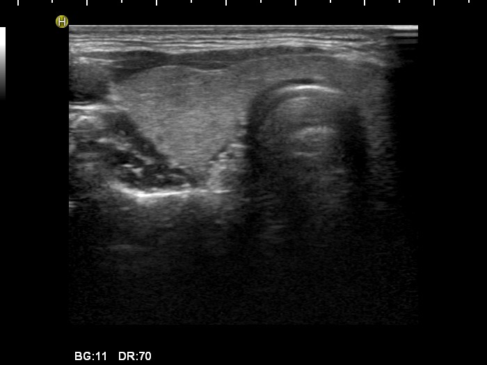
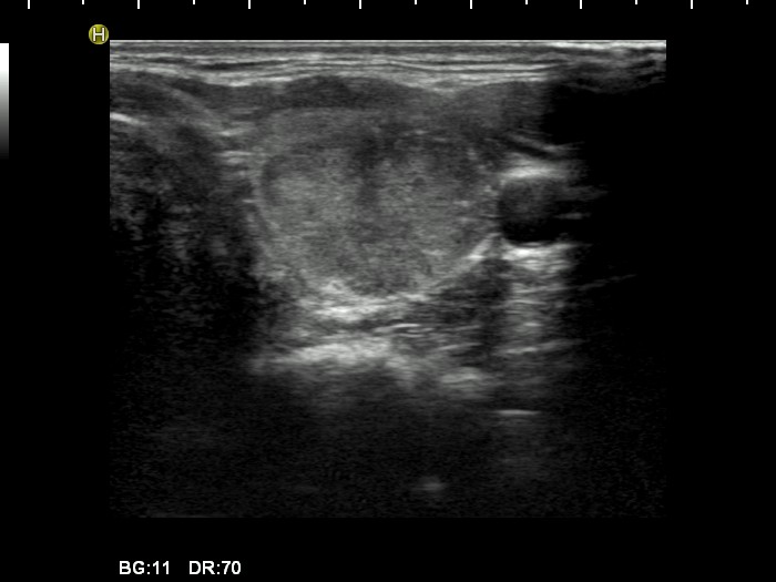
Ultrasonography. Tthe thyroid was echonormal. There was a hypoechogenic nodule in the ventral part of the left lobe. The borders of the nodule were irregular, the lesion contained microcalcifications and displayed an irregularly increased intranodular blood flow.
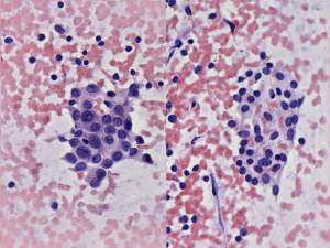
Cytological diagnosis: Papillary cancer. Lymphocytic thyroiditis.
Histopathology: Papillary cancer with focal lymphocytic thyroiditis. Metastases to the submandibular lymph nodes.
Comments:
-
The sonographic pattern is highly suspicious for papillary cancer. Most of the important sonographic properties specific for this type of cancer are presented: hypoechogenicity, irregular borders, microcalcifications, irregularly increased intranodular blood flow.
-
It is worth analyzing the cytological images - two cell populations were found. Firstly, normal thyroid follicular cells, secondly follicular cells displaying the features of papillary cancer.


