
100 consecutive patients with papillary carcinoma - Case 37. (ultrasonographic picture 4)

|

|

|
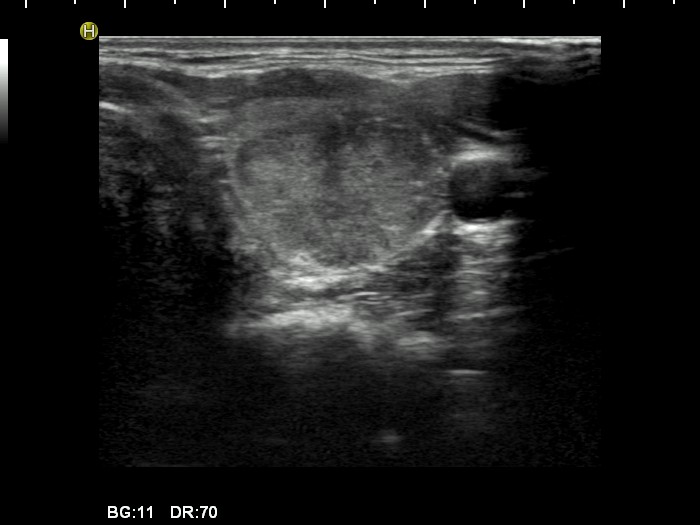
Left lobe, horizontal view. A moderately hypoechogenic nodule with several bright hyperechogenic granules. Note the irregularity of the ventral surface.


