
|
|
100 consecutive patients with papillary carcinoma - Case 42.
|

|


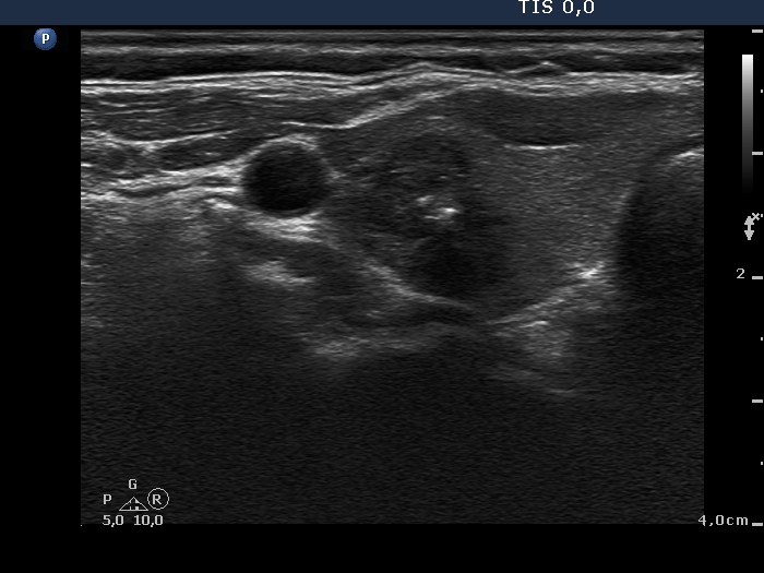
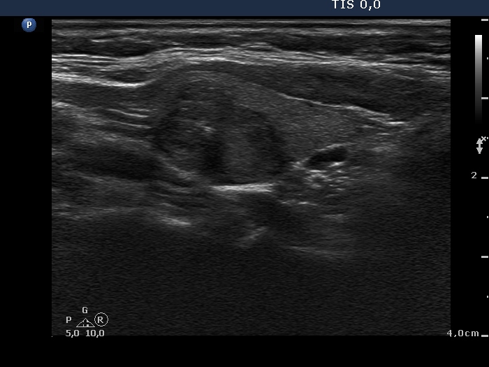


Clinical data: A 53-year-old woman was referred for evaluation of a nodular goiter detected on screening.
Palpation: The surface of the right lobe was uneven.
Functional state: euthyroidism (TSH 1.92 mIU/L).
Ultrasonography. The thyroid was echonormal. There was in the right lobe a hypoechogenic nodule, which presented irregular borders, hyperechogenic figures, increased intranodular blood flow and cystic degeneration.
Aspiration cytology was performed from the nodule and we gained a minimal amount of brown fluid. The cytological pattern itself was not reassuring.
We took the ultrasound presentation into account and gave a common ultrasound-cytological diagnosis of suspicion of papillary carcinoma.
Total thyroidectomy was performed. Histopathology disclosed papillary carcinoma.
Comment.
-
The cytological pattern itself was not enough to a definite carcinoma diagnosis. The lack of inclusion is a very rare phenomenon in the event of a papillary carcinoma. Moreover, there were only scattered number of nuclei with groove and there was no one typical papillary cluster on the smear. Although the nuclear crowding, the irregular chromatin structure and the mild pleomorphism are only weak signs of malignancy, the presence of all these features together are remarkable.
-
Combining the cytological data with the ultrasound presentation led to the combined ultrasound-cytological diagnosis.
- The interpretation of the intranodular hyperechogenic figures is not simple. On thorough analysis of the video, it seems more likely that these correspond to proliferation of connective tissue because of the simultaneous presence of bright lines and granules.


