
100 consecutive patients with thyroid nodule - Case 11. (ultrasonographic picture 4)
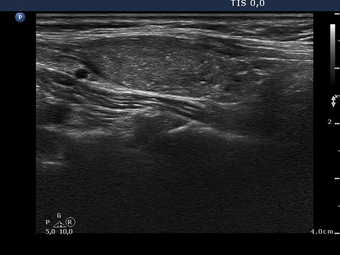 Lower part of the right lobe, another longitudinal scan. The lesion displays non-specific granules and more bright hyperechogenic figures. The latter are microcalcifications. |


