
100 consecutive patients with thyroid nodule - Case 25. (ultrasonographic picture 9)
|
|

|
|
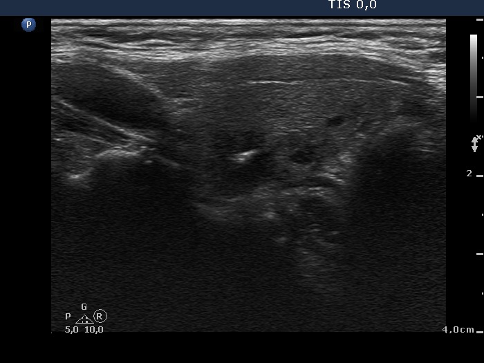
Left lobe, longitudinal scan. There is an echonormal lesion with cystic changes lower to the larger hypoechogenic lesion. The latter has irregular, ill-defined borders.