
100 consecutive patients with thyroid nodule - Case 25. (ultrasonographic picture 10)

|

|

|
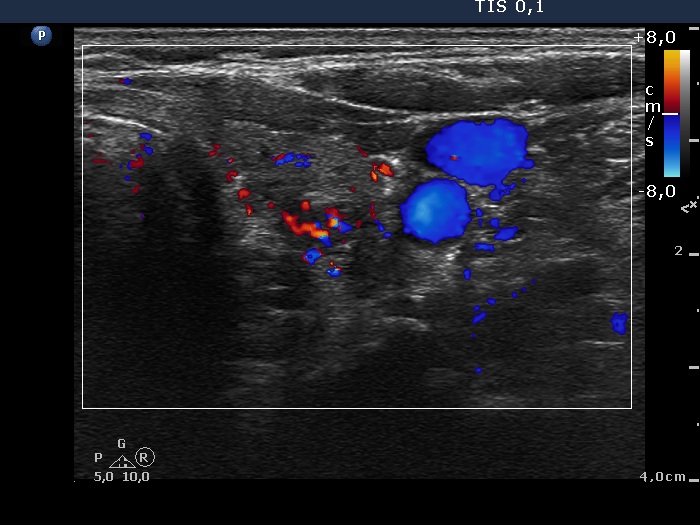
Left lobe, horizontal scan, color Doppler mode. The lesion presents a type 3 vascular pattern, i.e. increased intranodular blood flow.


