
QUICK TOUR - Case studies - sample case 4 of 6:Medullary carcinoma - Case 19. (ultrasonographic picture 7)
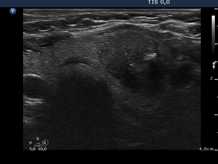 Lower part of the left lobe, horizontal scan. There is a hypoechogenic nodule presenting several microcalcifications and an irregular larger hyperechogenic figure. The latter corresponds to amyloid deposit. |


