
QUICK TOUR - Case studies - sample case 6 of 6:The role of complex diagnosis - follicular proliferation - Case 3. (ultrasound picture 7)
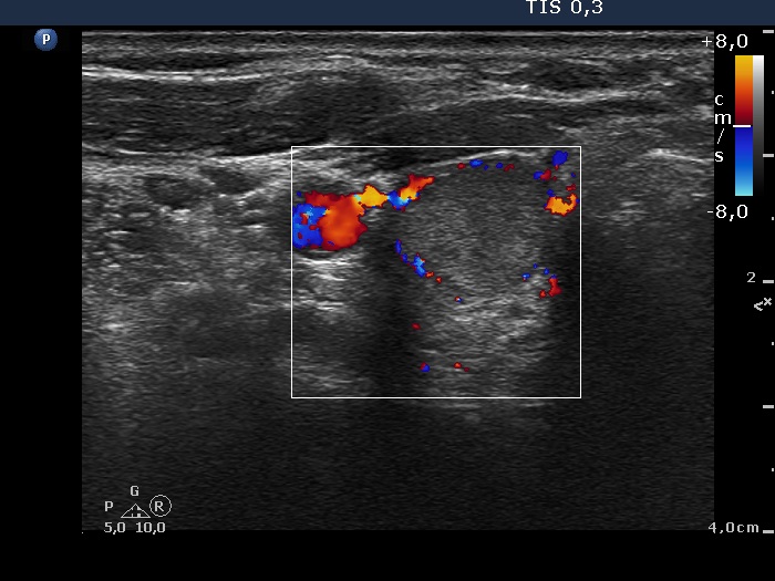 Lower pole of the right lobe, horizontal scan, color Doppler mode. This nodule displays a type 2 vascular pattern. |
