
QUICK TOUR - Case studies - sample case 6 of 6:The role of complex diagnosis - follicular proliferation - Case 3. (ultrasound picture 11)
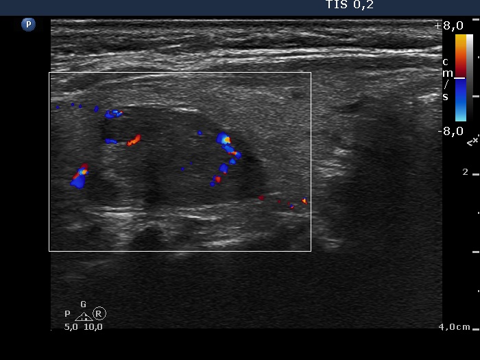 Left lobe, longitudinal scan, color Doppler mode. The largest nodule within the large nodular area presents a type 3 vascular pattern. |
