
QUICK TOUR - Case studies - sample case 5 of 6:
Secondary thyroid carcinomas - Case 15. (ultrasonographic picture 4)
Metastasis of a laryngeal adenoid cystic carcinoma
|
|

|
|
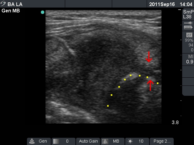
Left lobe, longitudinal scan. The hypoechogenic mass invades (red arrows) the echonormal part of the thyroid. The contour of the trachea is marked with yellow.