
|
|
The borders of the nodule - case 1527
|
|


Clinical presentation: A 61-year-old woman was referred for evaluation of thyroid nodules discovered on screening. She was told that the lesion in the left lobe is suspicious because of microcalcifications.
Palpation: a multinodular goiter with no suspicious nodule on palpation.
Hormonal examination: euthyroidism with TSH-level 0.83 mIU/L.
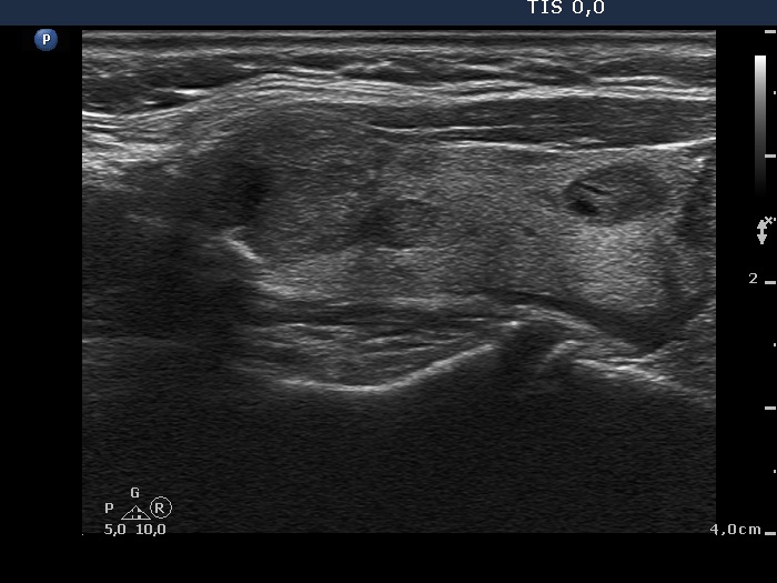
Ultrasonography. The thyroid was echonormal and contained numerous nodules with different echogenicities. A nodule in the upper pole of the right lobe and another one in the central part of the left lobe were relatively suspicious. The latter had irregular borders and presented perinodular blood flow and hyperechogenic granules.
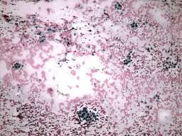
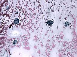
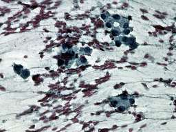
Cytology of the nodule in the upper pole of the right lobe resulted in benign colloid goiter, while the cytological pattern of the left nodule corresponded to follicular proliferation.
A combined ultrasound-cytological diagnosis was given in the case of the left nodule: benign follicular proliferation with less than 1% risk of carcinoma.
We advised regular follow-up instead of surgery. The patient decided to undergo on surgery.
Histopathology disclosed benign hyperplastic nodules.
Comments.
-
The ultrasound presentation of the whole thyroid corresponds to hyperplastic nodular thyroid disease. The lesion in question presented neither halo sign nor perinodular blood flow, therefore the risk of a follicular tumor was very low.
-
The cytological pattern is identical with that of a follicular tumor, which is not an infrequent finding in hyperplastic nodules. In around 30 to 50% of all cytologically diagnosed follicular tumors, histopathology discloses hyperplastic nodules.
-
The various nodules present lobulated and spiculated margins.