
|
|
The composition of the nodule - case 2104
|
|


First examination (first and second rows of images)
Clinical presentation: A 39-yr old woman was referred for evaluation of a thyroid cyst which has refilled twice in the past. On the last aspiration, 2 years ago the maximal diameter of the nodule was 24 mm.
Palpation: no abnormality.
Laboratory test: 2.71 mIU/L.
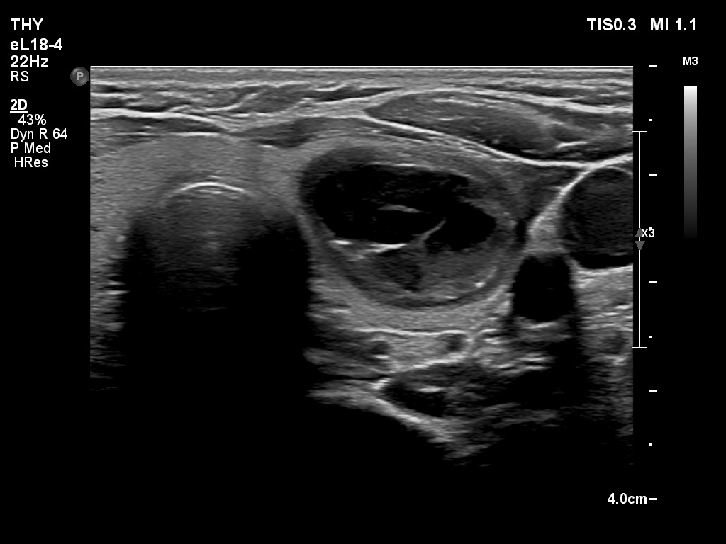
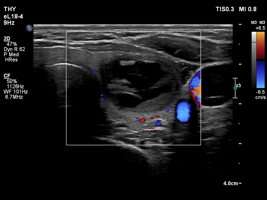
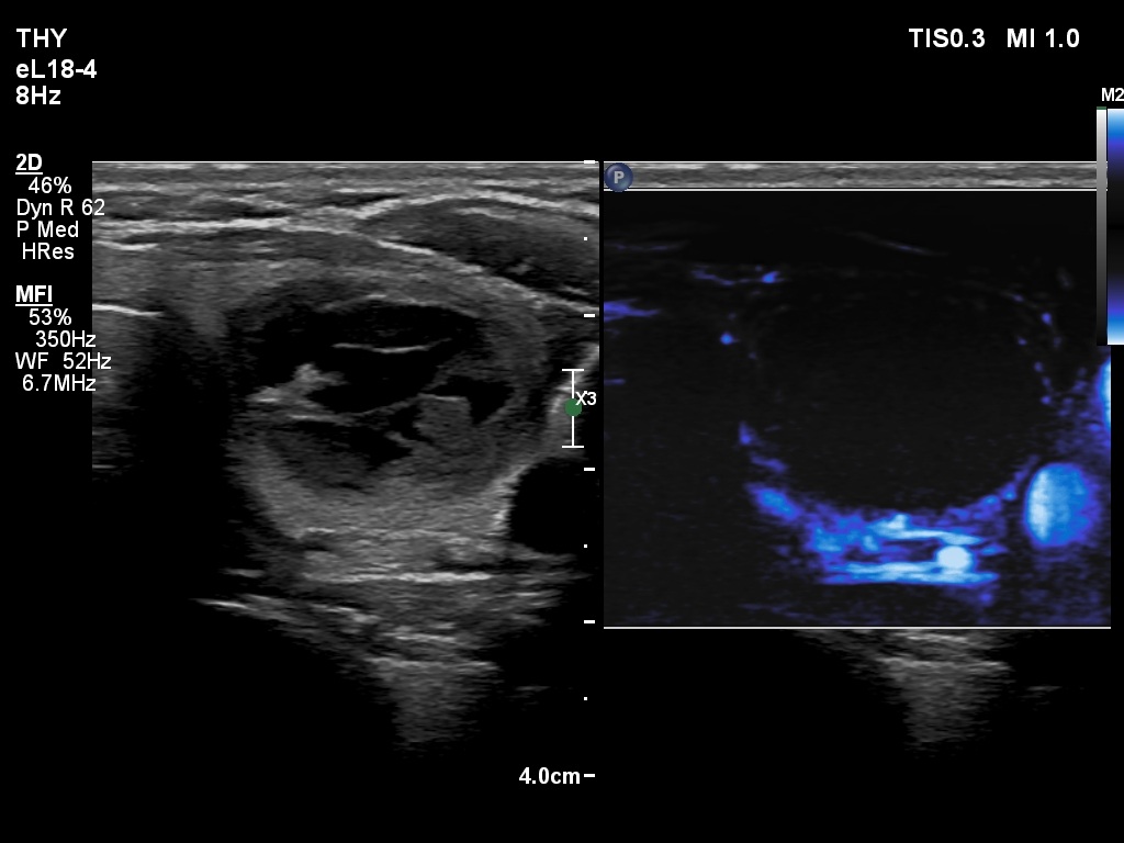
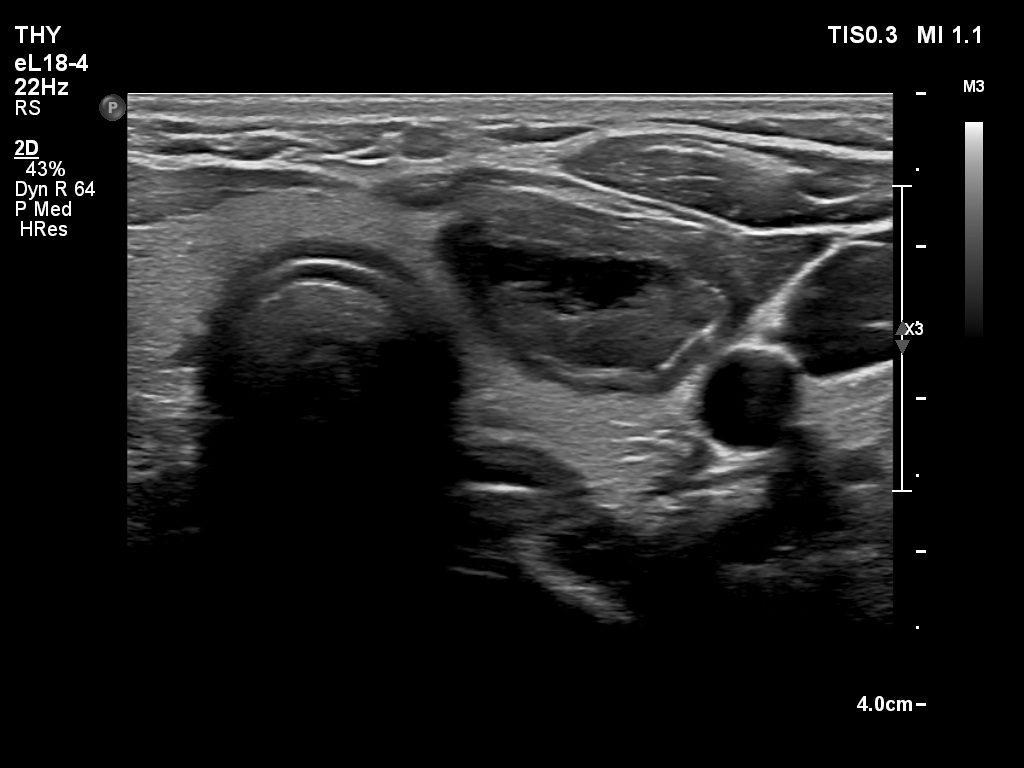
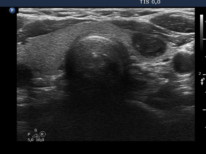
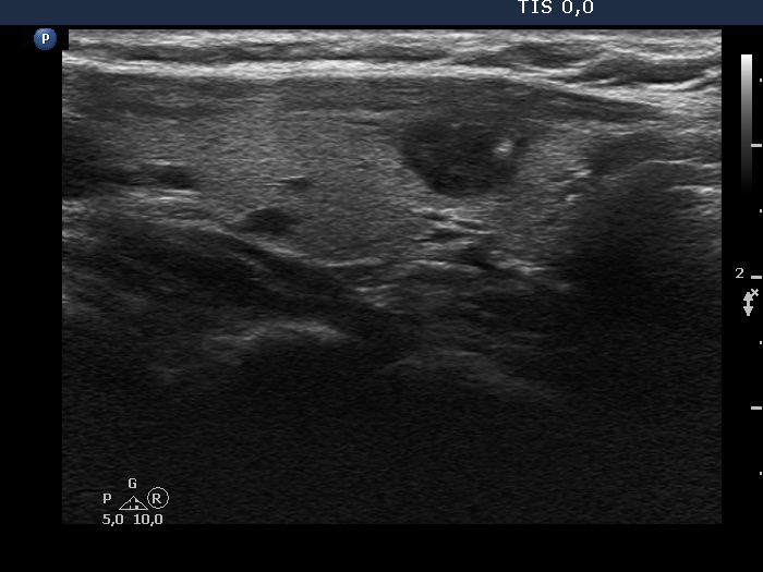
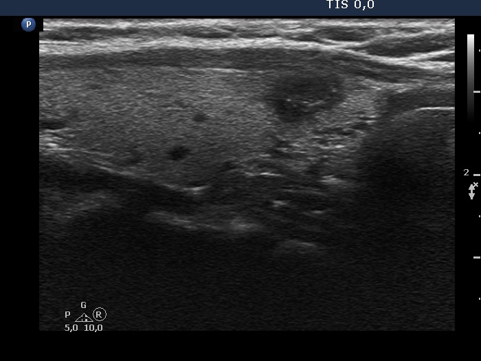
Ultrasonography. The thyroid was echonormal. There was a central-type cystic nodule in the left lobe. The solid portion was echonormal-minimally hypoechoic.Cytology was performed from the lesion in the left lobe and resulted in benign, cystic-colloid goiter.
Suggestion. Ultrasound in two years, in the event of complaints at once. If the cyst would recur ethanol sclerotherapy is advised.
Five months after the first examination (third row of images)
Clinical presentation: The patient had no complaints. She was referred for evaluation of an elevated TSH level.
Palpation: no abnormality.
Laboratory tests: 4.76 mIU/L, aTPO 0,6 U/mL.
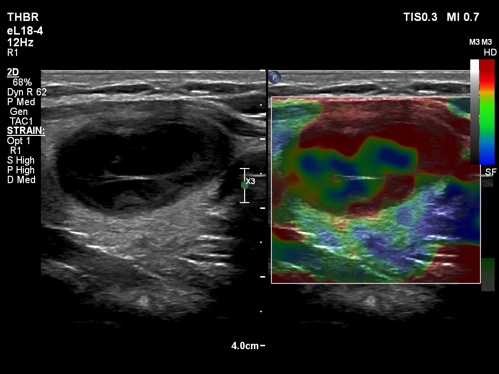
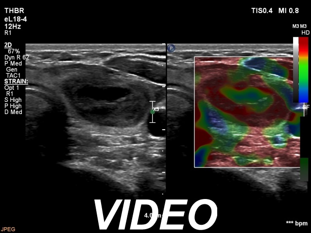
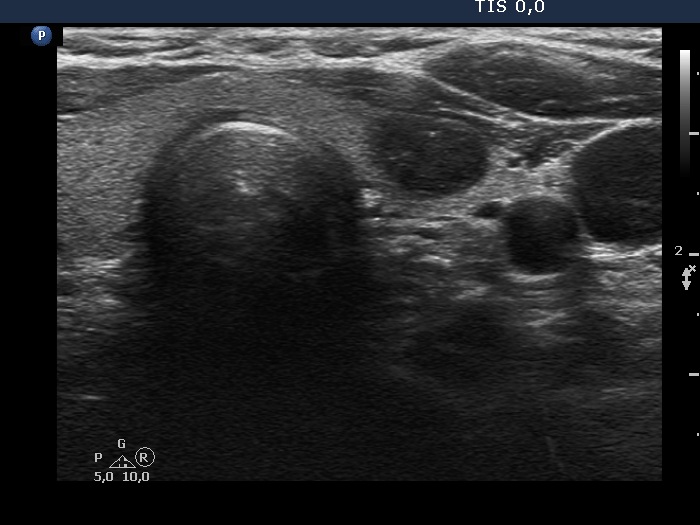
Ultrasonography. The thyroid was echonormal. There was a hypoechoic nodule in the ventral part of the left lobe. The ventral borders of the lesion were undefined because the echogenicity of the lesion and the neighboring strap muscle was identical.Suggestion: TSH in a year, ultrasound in two years, in the event of complaints at once.
Comments.
- Compared with the nodule just after the aspiration with that seen five months later, a spontaneous further decrease can be observed.
- On the second examination, we cannot see cystic portions, therefore the intranodular echogenic figures might cause concern.
- The presentation of the nodule at the follow-up is an example of non-pathological cause of blur.
- The normal value of the TSH is influenced by the BMI, lower the BMI higher the upper limit of the normal value.