
|
|
Case 974
|
|


Clinical presentation. A 33-year-old woman was referred for an evaluation of a goiter evolved over 6 months.
Functional state: hypothyroidism (TSH 21.8 mIU/L, FT4 8.32 pM/L).
Palpation: Both lobes were enlarged and very firm. No nodule was palpable.
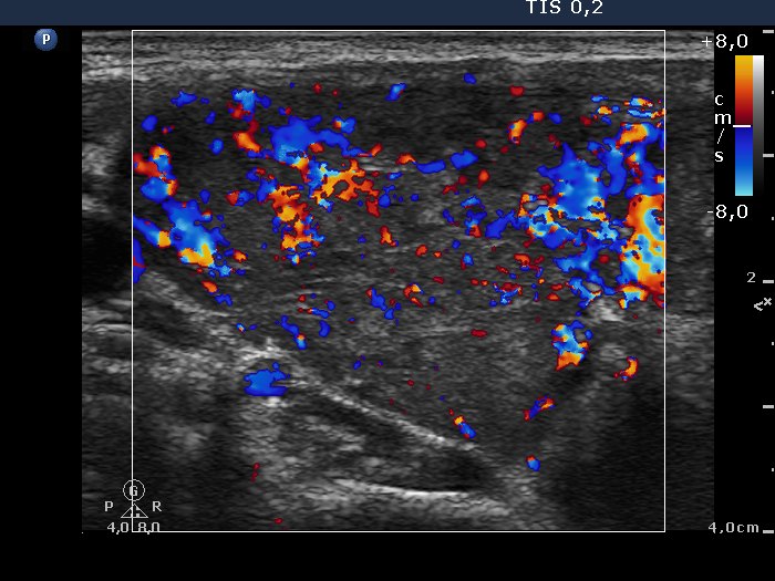
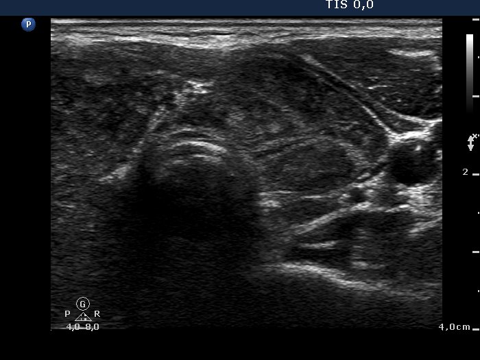
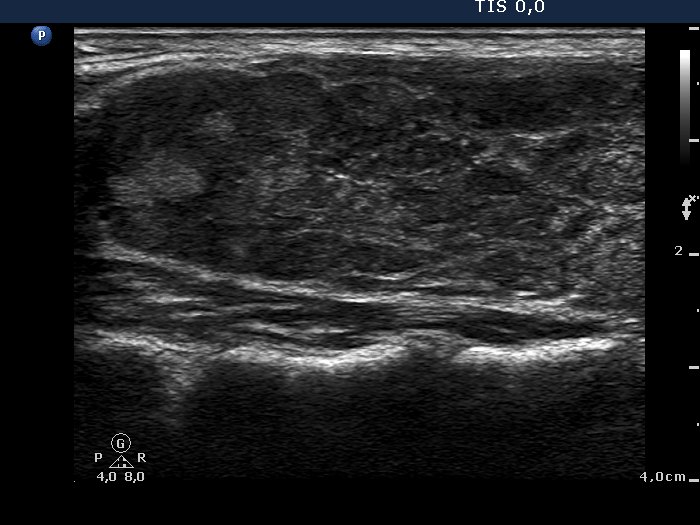
Ultrasonography: The thyroid was hypoechogenic and contained numerous discrete echonormal lesions divided by fibrous tissue. The vascularization was increased.
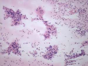
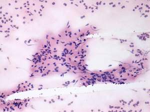
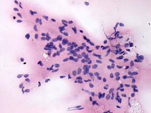
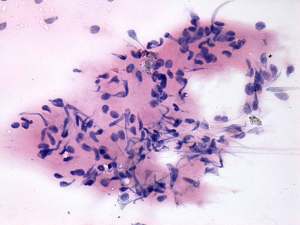
Cytology was performed and resulted in thyroiditis. There were scattered number of lymphocytes and multinucleated giant cells were also found.
Anti-TPO exceeded 900 U/mL.
Final combined diagnosis: primary hypothyroidism caused by autoimmune thyroiditis.
Comments.
-
The sonographic pattern corresponds to the so-called micronodular (or pseudonodular) form of lymphocytic thyroiditis.
-
The presence of multinucleated giant cells composed of epithelioid cells resembles that observed in de Quervain's thyroiditis. This type of multinucleated cells occurs rarely in autoimmune thyroiditis.