
Thyroid cancers - case 908 (cytologic picture 2 of another smear)
|
|

|
 |
|
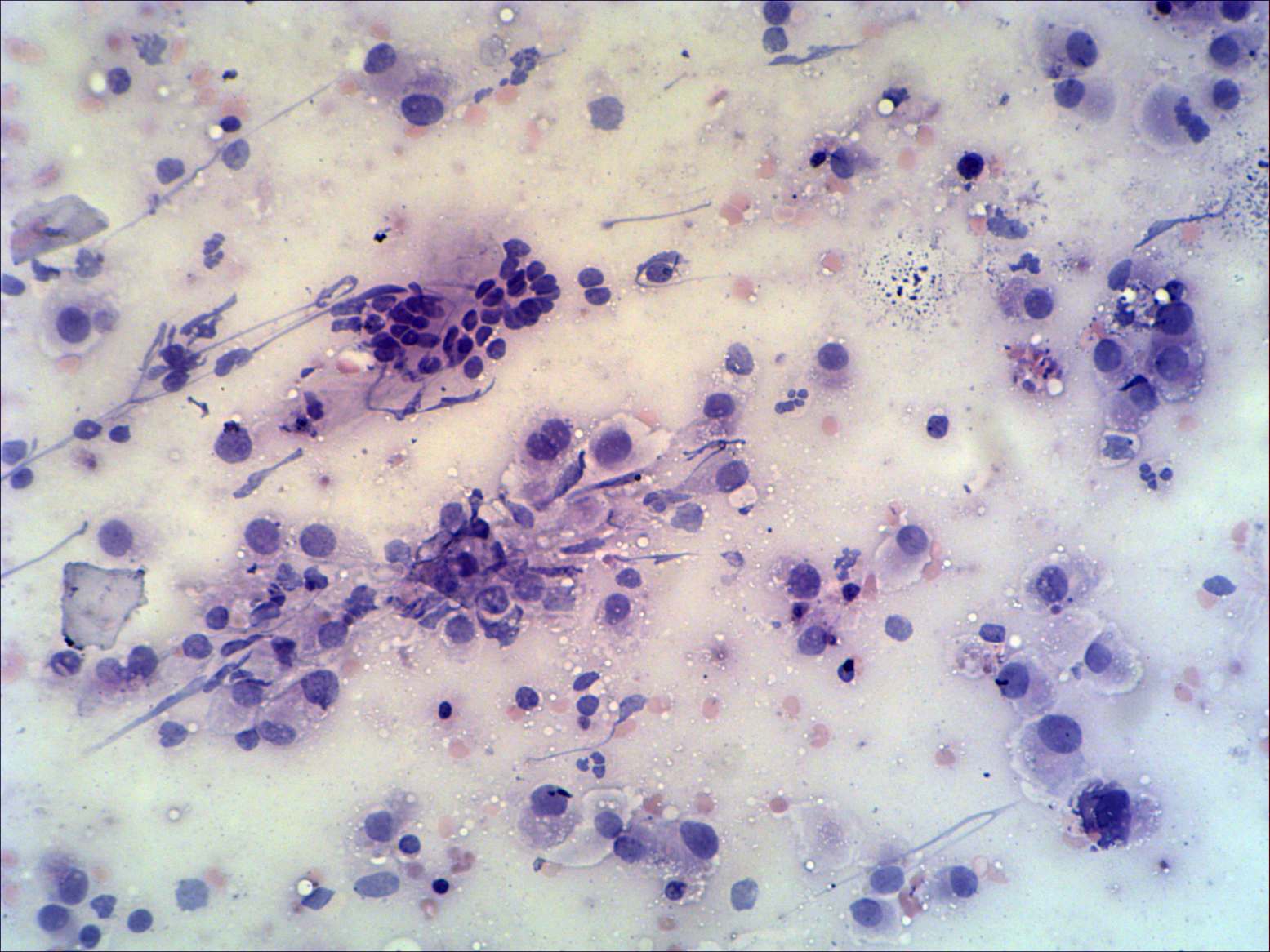
Pap-staining, 200x. Beside enlarged oxyphilic cells, there is a cell group composed of non-metaplastic cells in the center of the image. Note nuclear debris. This pattern is identical to that observed in the event of a Hashimoto thyroiditis.