
|
|
Thyroid cancers - case 908
|
|


Clinical data: A 61-year-old woman was referred for evaluation of a rapidly growing mass in the right thyroid. The tumor developed within 5 weeks..
Palpation: a very hard, not freely moveable nodule in the moderately enlarged right lobe. Multiple nodules were palpable in both lobes.
Functional state: euthyroidism (TSH-level 0.48 mIU/L).
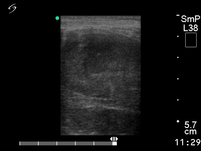
Ultrasonography. The basic echo structure of the thyroid was normal. There were multiple hypoechogenic nodules in the right lobe.
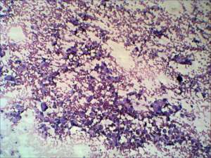
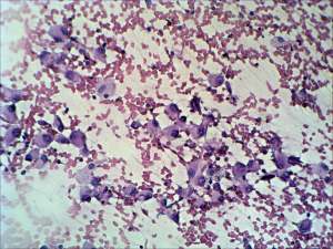
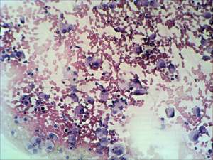
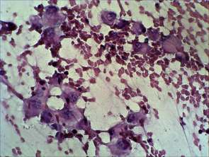
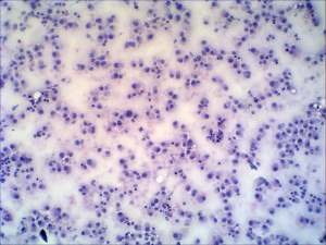
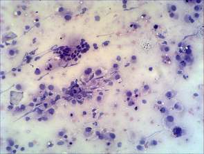
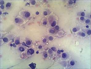
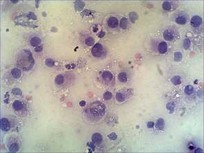
Cytological picture.Tthere was no colloid in the background. Thyrocytes occurred mostly dissociated. Great proportion of follicular cells exhibited oxyphilic metaplasia. The nuclei presented moderate degree of anisonucleosis and several cells had intranuclear inclusions. Otherwise, the cytological pattern was identical to that observed in a Hürthle-cell tumor. Cytological diagnosis: oxyphilic tumor with higher than the average risk for invasive tumor.
Combined clinical-cytological diagnosis: thyroid cancer, possible anaplastic cancer.
Histopathology: anaplastic cancer.
Comment. This is an unusual cytological pattern because of the relatively reassuring pattern and the lack of pleomorphic cells on the smears. The cytological pattern is in sharp contrast with the clinical picture.
It is likely that the smear was gained from the more differentiated part of the tumor.
.