
|
|
100 consecutive cases of papillary cancer - case 080
|
|


Clinical data: A 36-year-old woman was referred for evaluation of a thyroid nodule. Despite benign cytological report, the endocrinologist held the nodule suspicious.
Palpation: a firm nodule in the upper part of the left lobe.
Laboratory test: TSH 0.91 mIU/L.
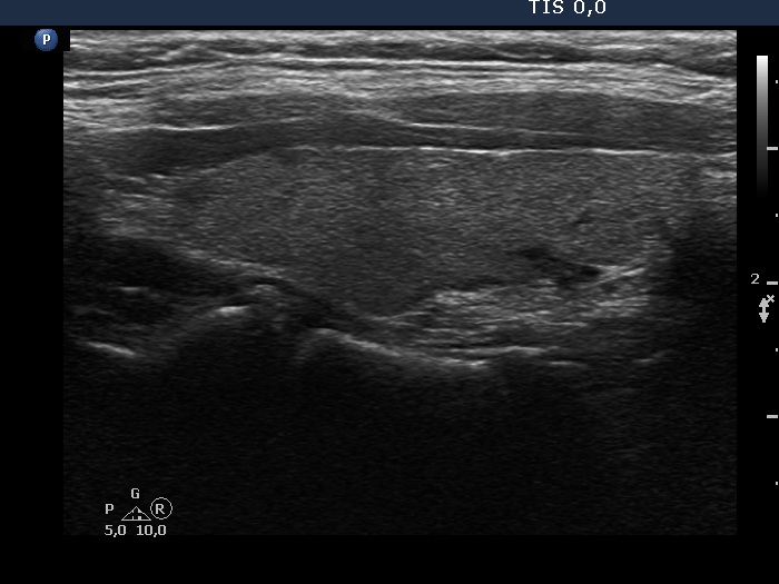
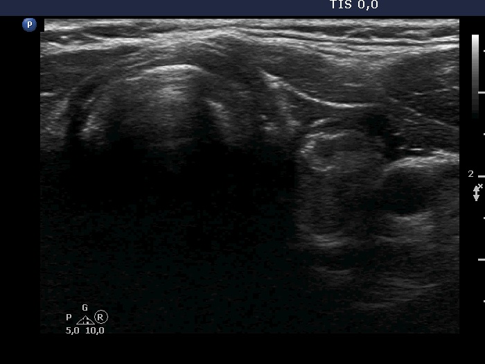
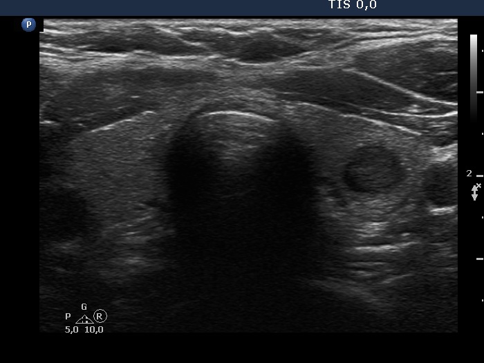
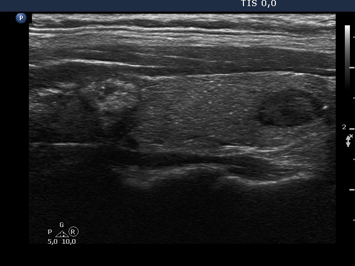
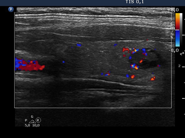
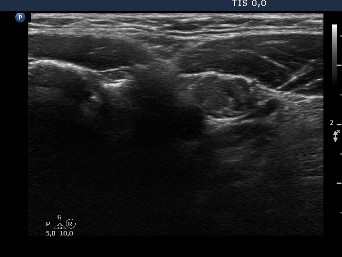
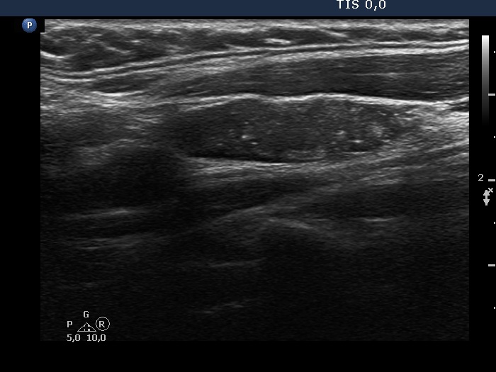
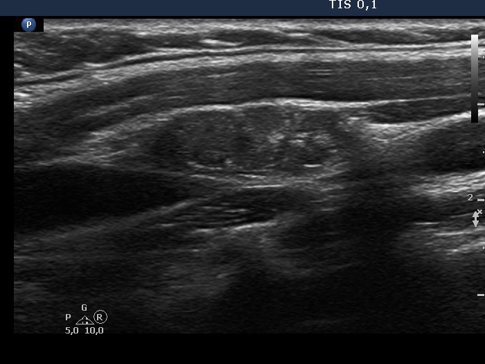
Ultrasonography. The thyroid was echonormal. There were two nodules in the left lobe. The upper lesion was a dominantly isoechoic nodule which had microcalcifications while the lower one was a hypoechoic nodule. There were numerous microcalcification relatively far from the primary tumor focus. The upper nodule was avascular while the lower one presented both intranodular and perinodular blood flow.
A conglomerate of lymph nodes was found 2 cm above the left lobe. The nodes did not have hilum but contained microcalcifications and presented irregular vascular pattern. (This was not noticed on the previous ultrasound examination.)
Cytology performed from the heterogeneous lesion and from the lymph node resulted in papillary cancer and metastasis of papillary cancer, respectively.
Total thyroidectomy was performed. Histopathology disclosed a multifocal papillary cancer with metastasis to the neck lymph nodes.
Comment. This case illustrates a very important role of thyroid ultrasound which is less emphasized in the literature. There are ultrasound patterns which still raise suspicion even if the cytology is benign. This pattern is suspicious enough to indicate surgery even when the presence of pathological lymph nodes would be ignored.