
|
|
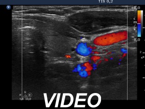
100 consecutive cases of papillary cancer - case 081
|
|


Clinical presentation: A 42-year-old woman requested an examination. She was diagnosed with a nodules goiter 12 years ago. Cytology was performed twice in other hospitals, first with a benign and second with a non-diagnostic repost, 12 and 3 years ago, respectively. In the last few years, the patient felt a rapid increase in nodular lesion.
Palpation: a firm nodule in the lower pole of the right lobe.
Laboratory tests: TSH 2.22 mIU/L, aTPO below 28 U/mL.
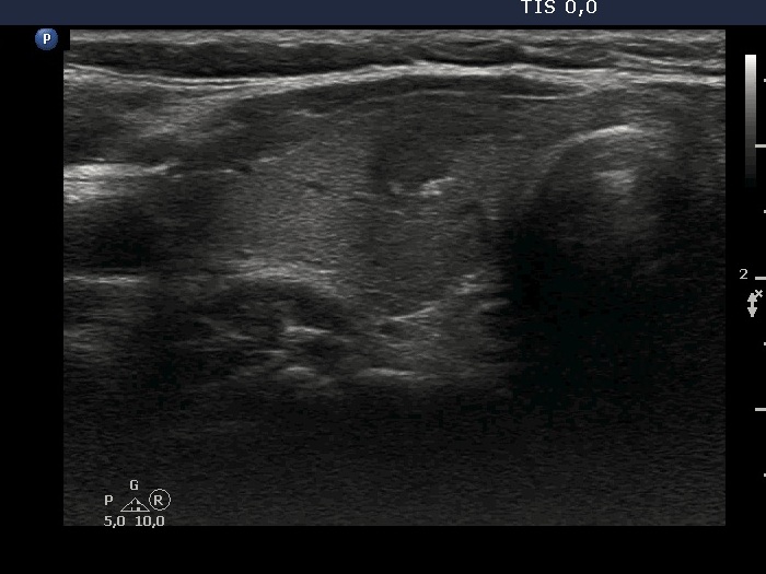
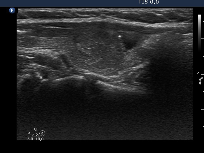
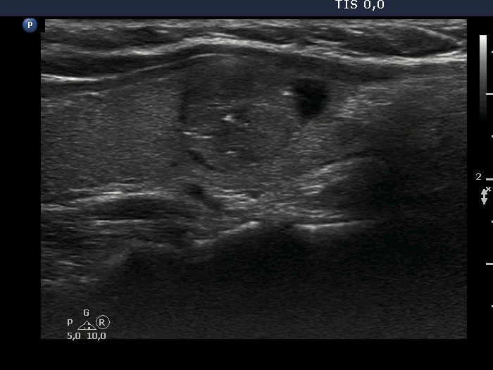
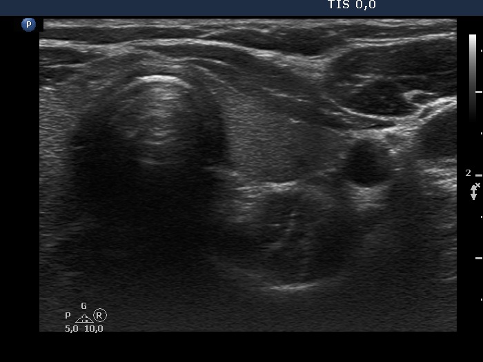
Ultrasonography. The thyroid was echonormal or minimally hypoechoic. There was a moderately hypoechoic nodule which had a cystic portion in the lower part. The nodule had lobulated margins and contained different hyperechoic figures. Beside figures caused either by posterior enhancement of proliferation of connective tissue, several ambiguous. bright granules were also found. Compared with the first measurement, the nodule is increased by more than 75% in volume.
Cytology resulted in suspicion of papillary cancer.
Histopathology revealed a papillary cancer which infiltrated the capsule of the gland. Growth extrathyroidal extension was not detected.
Comment. It is worth looking at the video several times and analyzing the various echogenic foci.