
|
|
Introduction - case 150
|
|


Clinical presentation: A 60-yr-old woman was referred for evaluation of a thyroid nodule detected on routine medical examination.
Palpation: a large firm nodule in the right lobe.
Laboratory tests: TSH 5.47 mIU/L, aTPO 172 U/mL.
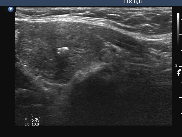
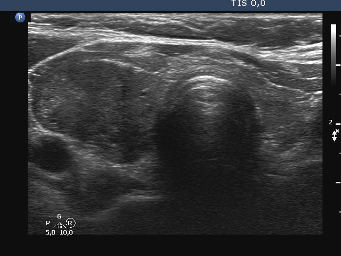
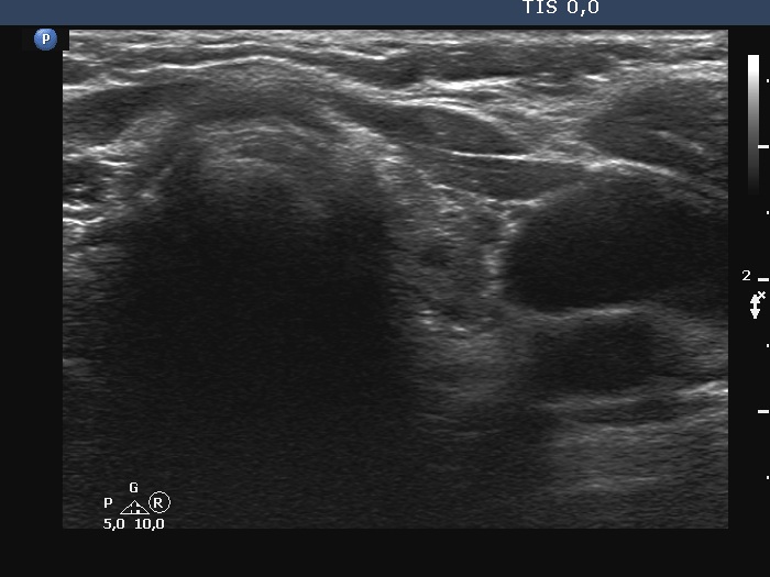
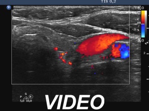
Ultrasonography. The right lobe was substantially enlarged and hypoechoic. It had macrocalcifications. The issue was whether the lobe was composed of a large nodule or only diffuse enlargement was present. The left lobe was much smaller. This lobe was also hypoechoic but to a lesser extent as was the right lobe. The vascularity was increased in the right while decreased in the left lobe.
FNA of the nodule resulted in lymphocytic thyroiditis.
Comment.
-
The issue was whether the lobe was composed of a large nodule or only diffuse enlargement was present. On some still images, it seemed evident that there was a nodule. But we should never forget, that the reality is much more represented by videos. When we stop the continuous scan significantly affects what we see in a still image, which can therefore very easily be misleading. In the video, I highlighted those features on which we could decide considering only the ultrasound presentation that this very likely a nodule.
- The most decisive fact was the palpation that this was a nodule.