
Introduction - case 193 (ultrasonographic picture 3)
|
|

|
|
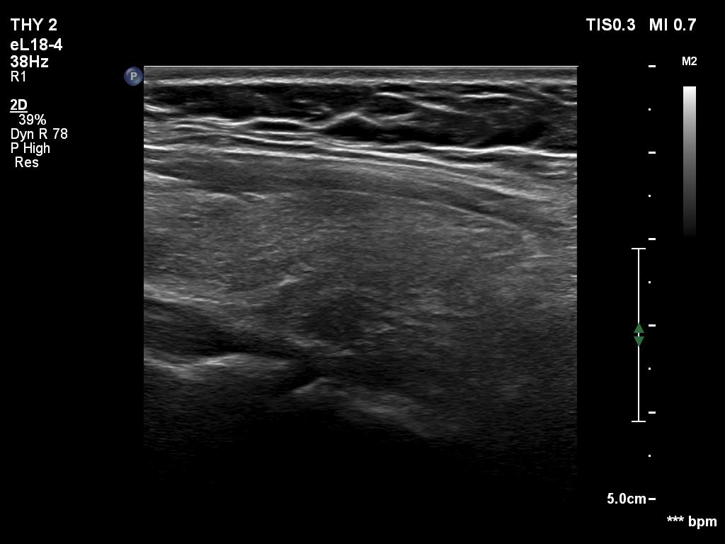
Right lobe, longitudinal view - set to higher frequency (better resolution). In this image, the presence of a hypoechoic lesion seems to be very likely.