
|
|
Introduction - case 193
|
|

Clinical presentation: A 19-yr-old, overweighted woman (161 cm height, 91 kg weight) was referred for aspiration cytology of a nodule. Neck and thyroid ultrasound were performed because of repeated upper airway infections.
Palpation: no abnormality.
Laboratory tests: TSH 2.12 mIU/L.
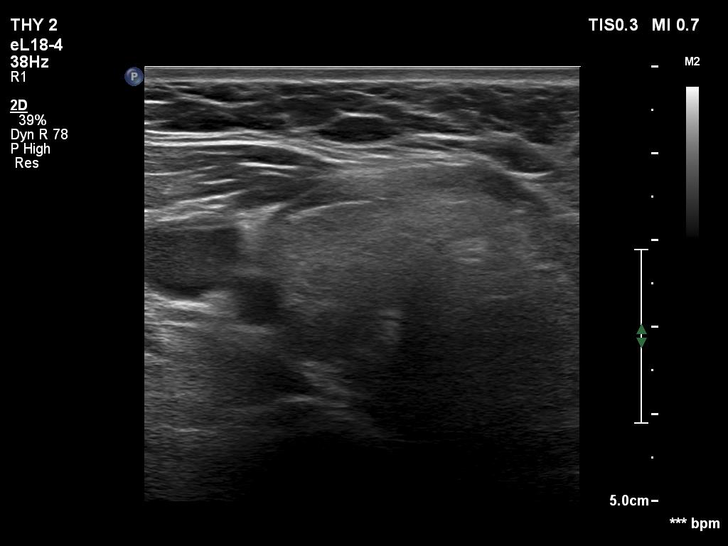
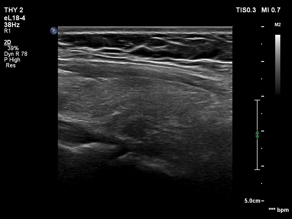
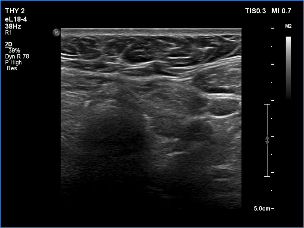
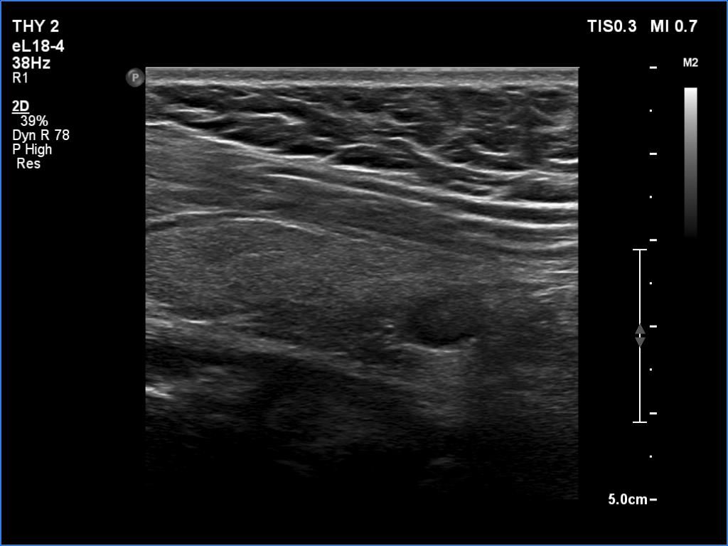
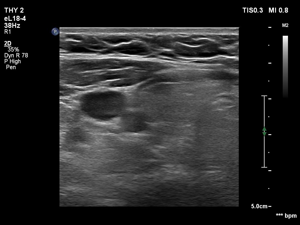
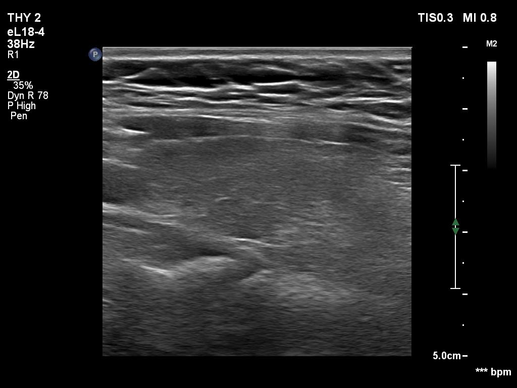
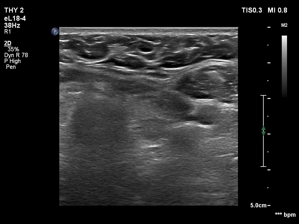
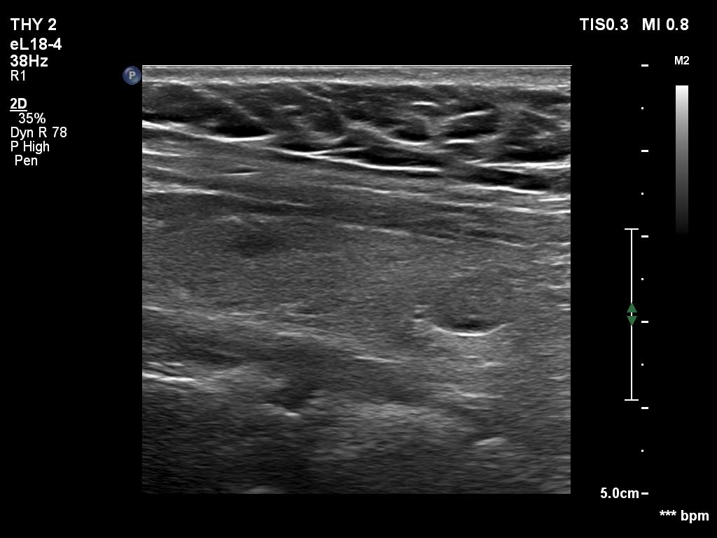
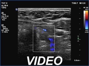
Ultrasonography. The thyroid was echonormal. Using higher frequency three discrete hypoechoic lesions were found, one in the middle dorsal part of the right lobe, one in the middle and a third one in the lower dorsal part of the left lobe. By decreasing the frequency, the presence of only the third one could be confirmed.
FNA of the nodule in the lower dorsal part of the left lobe resulted in benign colloid goiter.
Comment. It is a rule that larger the distance from the probe worse the permeability of ultrasound wave. This can lead in certain patients (overweighted ones or frequently in men) that the dorsal part of the thyroid lobe can deceptively look darker, i.e. hypoechoic. If we have any doubt, by lowering the frequency i.e. increasing the penetrance, the real situation can be much better to judge.
This happened in this patient. If we would trust the pattern gained by higher frequency settings, we would falsely diagnose at least one additional nodule.