
Introduction - case 193 (ultrasonographic picture 8)
|
|

|
|
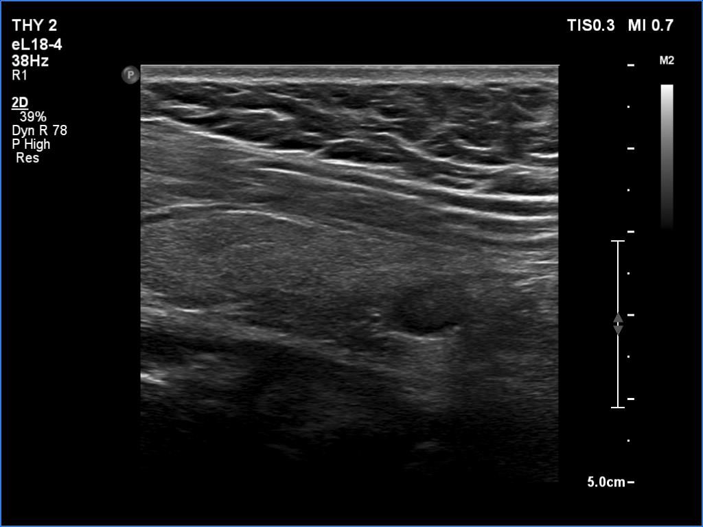
Lower part of the left lobe, longitudinal view - set to higher frequency (better resolution). Beside the nodule in the lower dosal part, a moderately hypoechoic area is also seen in the middle part of the lobe.