
|
|
Introduction - case 487
|
|


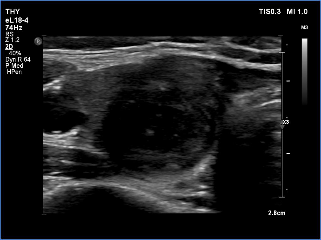
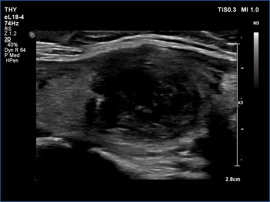
Clinical presentation: A 41-year-old woman was referred for evaluation of a nodule discovered on routine physical examination.
Palpation: The right lobe had a moderately firm nodule.
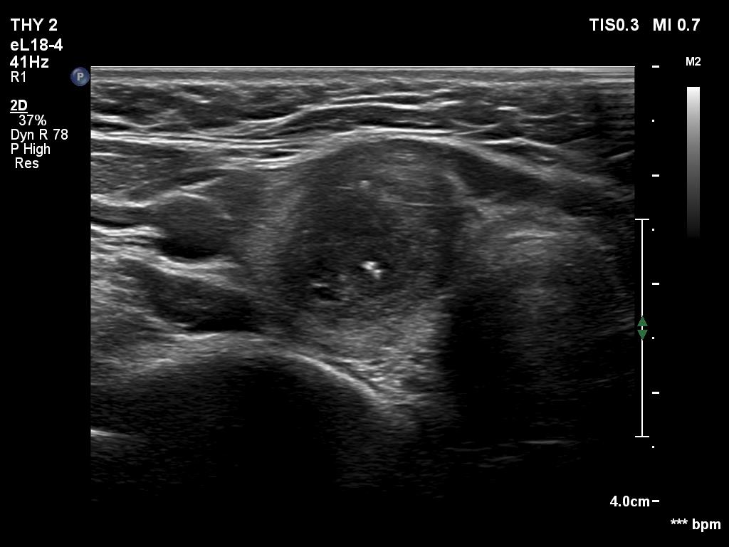
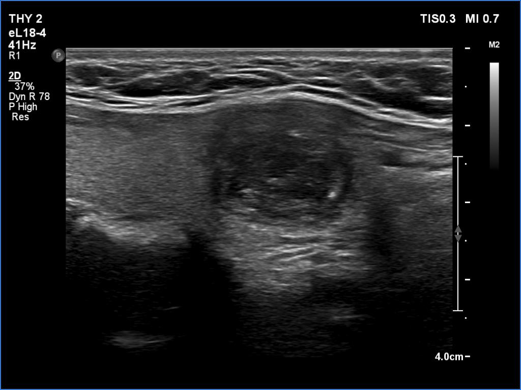
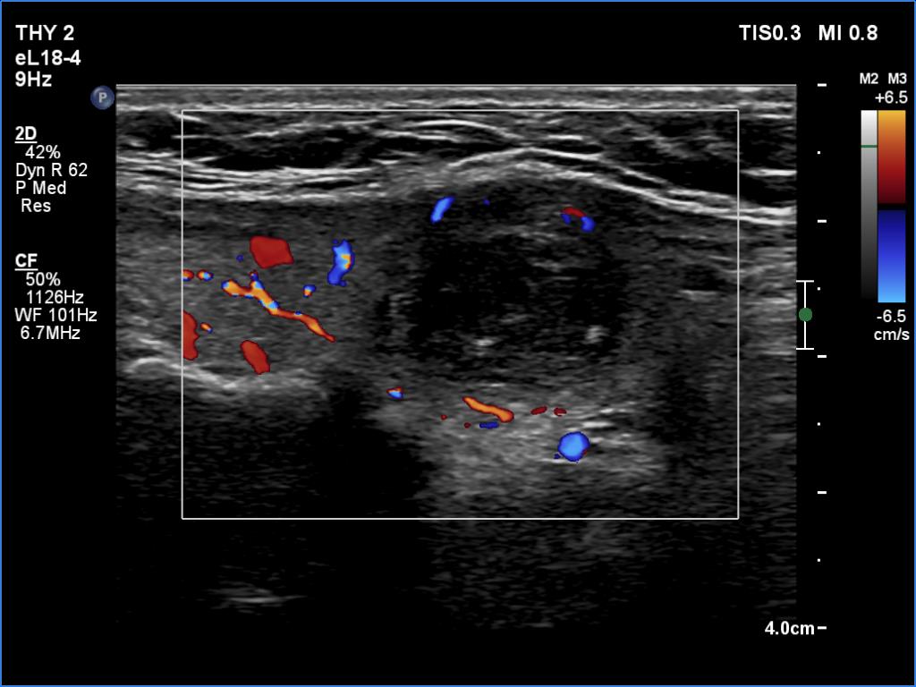
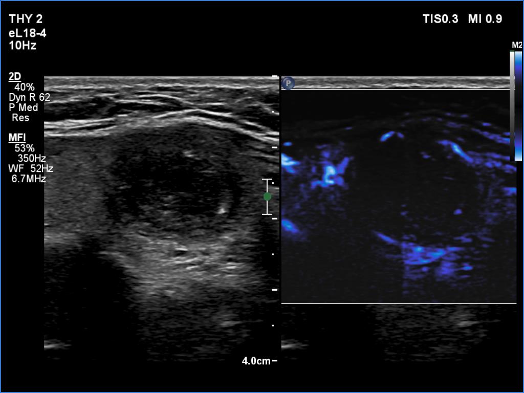
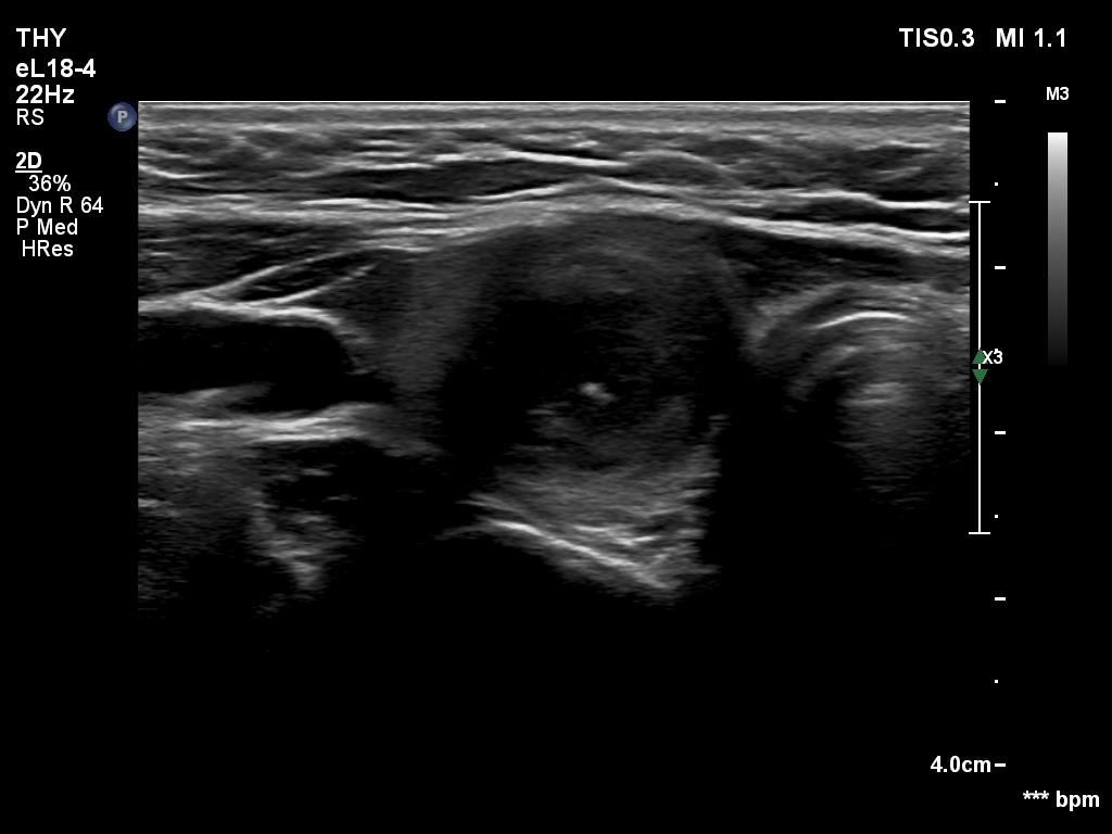
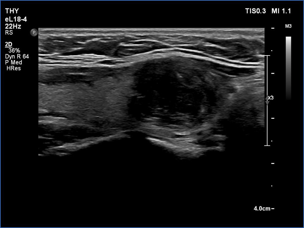
Ultrasonography revealed an echonormal thyroid. There was dominantly moderately hypoechoic nodule in the right lobe. Some parts of the lesion were deeply hypoechoic, and a few small cystic chambers were also within. The nodule presented with irregular shape and borders and had intranodular echogenic figures, primarily granules.
Laboratory tests: TSH 1.78 mIU/L, FT4 15.4 pM/L, anti-TPO 37 U/mL.
Aspiration cytology disclosed papillary cancer.
Total thyroidectomy was performed, histopathology resulted in T1b papillary cancer and nodular hyperplasia in the non-tumorous part of the thyroid.
Comment.
-
It is ambiguous how to interpret the echogenic figures. Although the presence of irregular shape and borders increases the likelihood that the echogenic granules are indeed microcalcifications, comet-tail artifacts must be also considered.
-
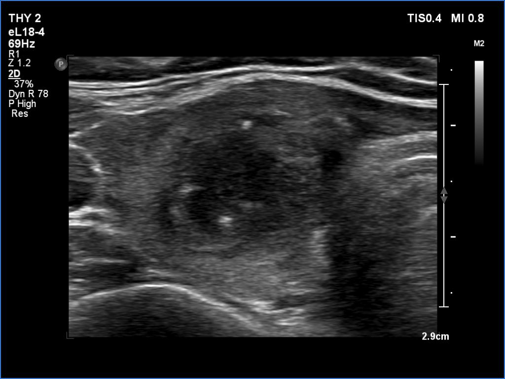
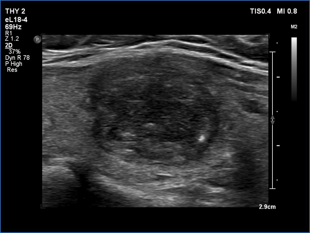
It is worth comparing the images recorded by using different settings. By using harmonization, we lose the details of very hypoechoic areas. Essentially, the latter seem to be anechoic.