
Introduction - case 462 (ultrasonographic picture 2)
|
|

|
|
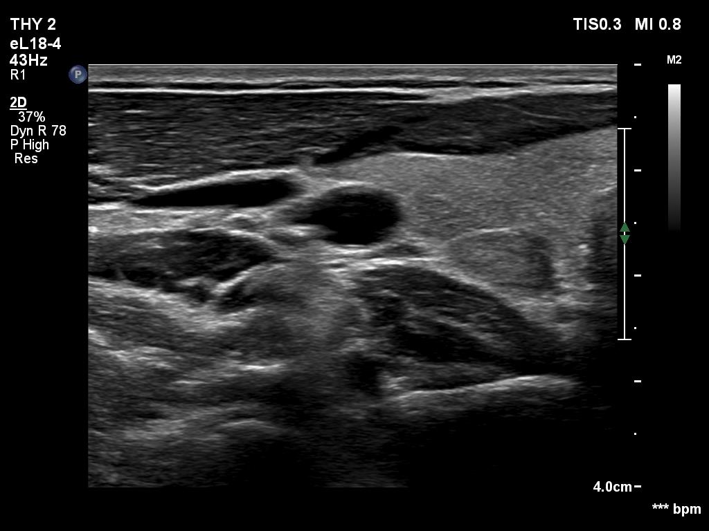
Right lobe, another transverse scan. The connective tissue band ventral to the minimally hypoechoic area hinders the penetration of ultrasound wave which makes the dosal tissue darker. Moreover, medial to the lesion (right in the image) a hypoechoic field can be seen. These artifacts make the appearance of the area deceptively nodular.