
|
|
Introduction - case 484
|

|


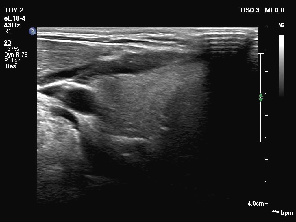
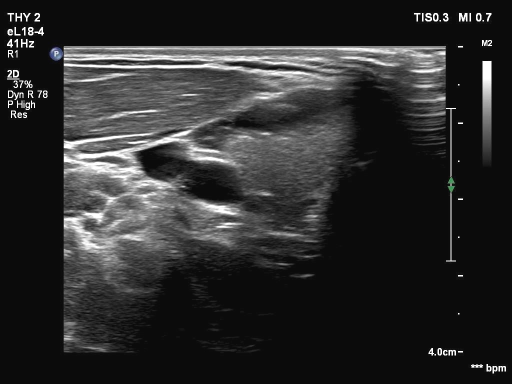
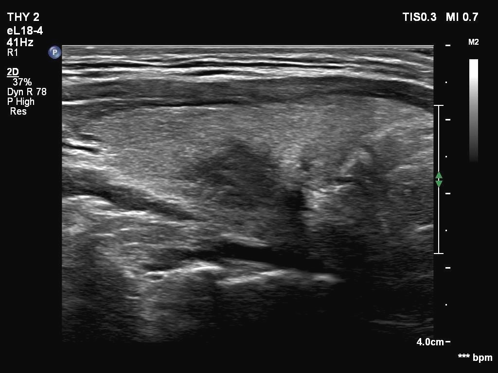
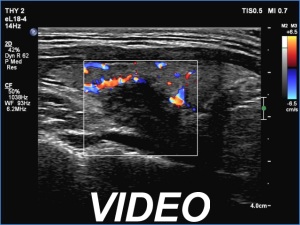
Clinical presentation: A 22-yr-old man was referred for ultrasound examination. He had 'lump in the throat feeling'.
Palpation: no abnormality.
Laboratory tests: TSH 0.83 mIU/L.
Ultrasonography The thyroid was echonormal. In certain longitudinal section, a hypoechoic discrete area had appeared in the dorsal region of the right lobe. Indeed, this was a tissue dorsal to the thyroid and probably corresponded to muscle fiber. Dorsal to this area, the esophagus could be identified.
Comment.
-
In most cases, we can place the transducer in such a way that parts of the thyroid gland appear nodular. Not infrequently, we can see nodule-looking areas without manipulation. As a rule, analysis of the video is always superior to making judgement on still images.
-
In this patient, dorsal to the hypoechoic area, the esophagus could be identified. This is quite a rare finding, in most cases the esophagus runs dorsal to the left lobe.


