
|
|
Introduction - case 816
|
|


Clinical presentation: A 9-yr-old boy was referred for evaluation of a suspected nodule. He was examined because of diffuse complaints which raised the possibility of thyroid disfunction. On ultrasound, an ambiguous discrete lesion was described.
Palpation: no abnormality.
Laboratory tests: TSH 4.23 mIU/L, aTPO 8 U/mL.
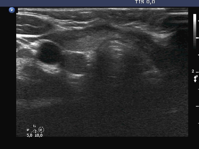
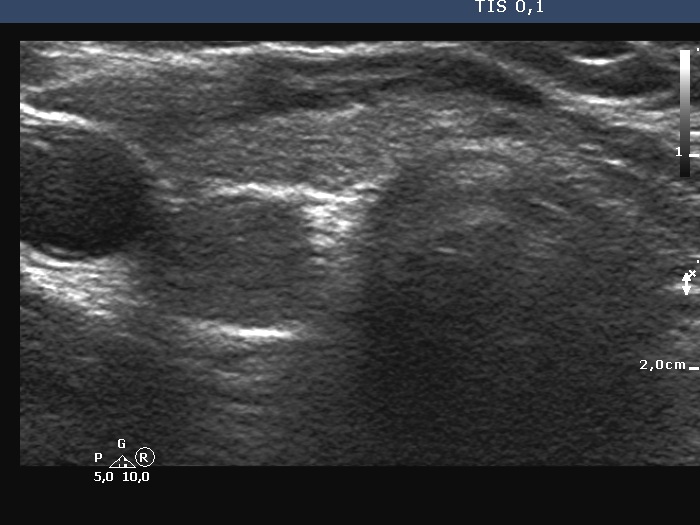
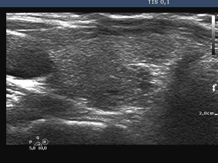
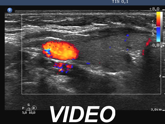
Ultrasonography. The thyroid was echonormal and had some minimally hypoechoic areas. A thick connective tissue band crossed the right lobe. Dorsal to this fragment the thyroid was very hypoechoic. On transverse scan, this area seemed to be a nodule. However, analysis of multiple sections revealed that this area was not a true nodule.
Comments.
-
The echogenicity of a specific tissue is deeply influenced by ventral structures. The well-known examples are the cystic fluid and the macrocalcification. The first can cause acoustic amplification, i.e. the dorsal structures become lighter, more echoic, while the macrocalcification has the opposite effect, the so-called acoustic shadowing makes the dorsal tissue very hypoechoic.
In this boy, the thick connective tissue hindered the penetration of the ultrasound wave, therefore the dorsal structure became darker, hypoechoic. -
The longitudinal scanning is almost always more important than the transverse investigation, because larger parts of the thyroid are visualized in the former.