
|
|
Introduction - case 960
|

|


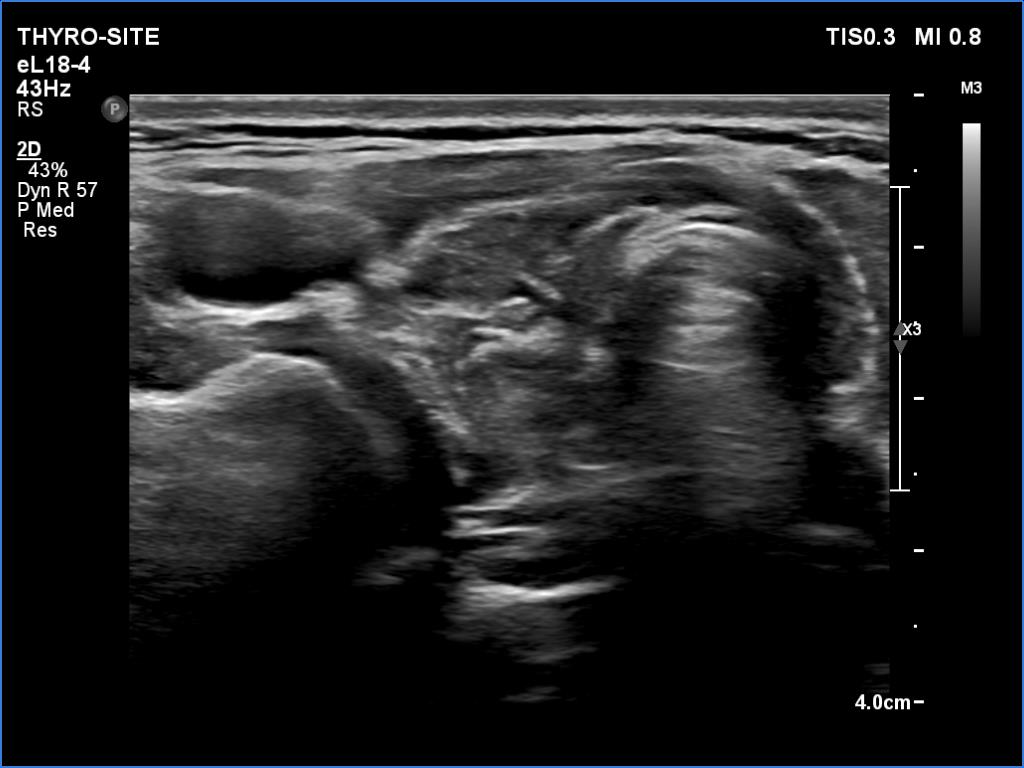
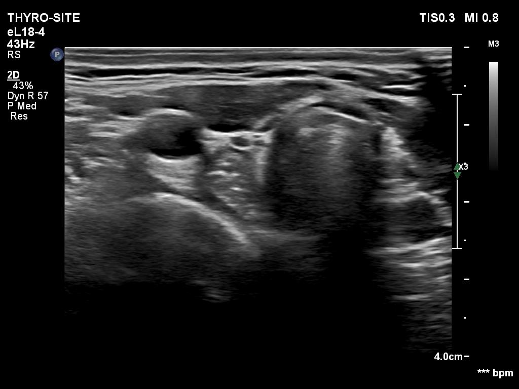
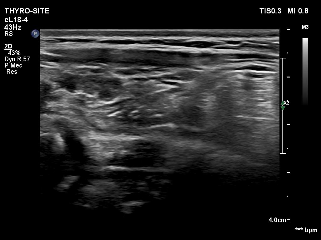
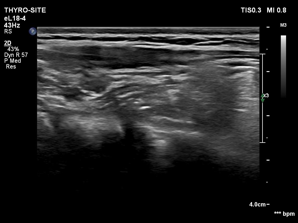
Clinical presentation: A 40-yr-old woman came to a routine follow-up. She has been treated for hypothyroidism for more than 15 years.
Palpation: no abnormality.
Laboratory test: TSH 0.91 mIU/L on daily 125 microgram levothyroxine.
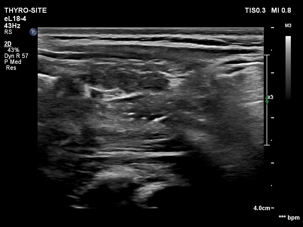
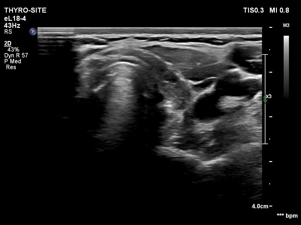
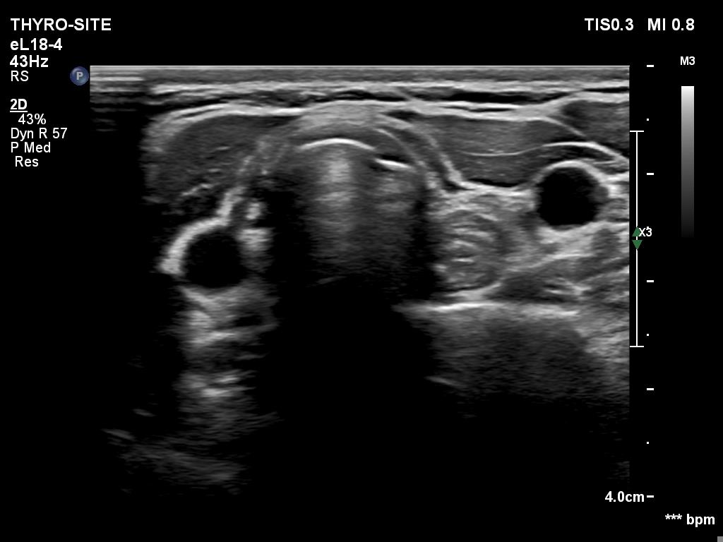
Ultrasonography. The thyroid was atrophic and hypoechoic. There were no discrete lesions within. The esophagus could be seen dorsal to both lobes.
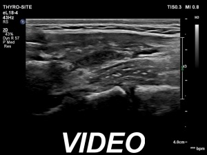
Comments. It is a rare finding that the esophagus can be seen also dorsal to the right lobe. The clue of the identification of the esophagus is the pipe-like running upwards and downwards. This can be seen in section parallel with the esophagus, i.e., in longitudinal scanning. In perpendicular sections, the presentation of the esophagus is very similar to a hypoechoic nodule presenting back wall figures.


