
|
|
The borders of the nodule - case 2107
|
|


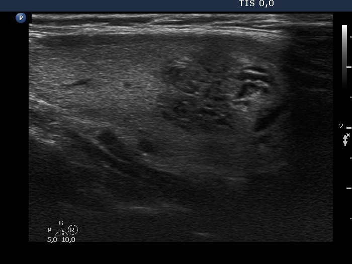


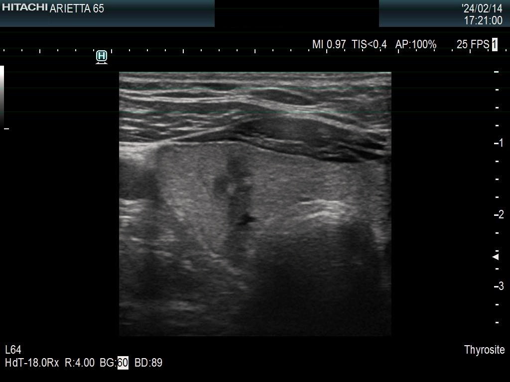

First examination (first row of images):
Clinical presentation: A 39-yr-old woman was referred for aspiration cytology. She has received thyrostatic therapy for two years. The nodule was detected on routine follow-up.
Palpation: There was a not firm nodule in the right lobe.
Laboratory tests: TSH 0.91 mIU/L, FT4 13.4 pM/L on daily 10 mg methimazole.
Ultrasonography. The thyroid was echonormal. There was an echonormal nodule in the middle of the right lobe. The nodule had intranodular echogenic lines. The dimensions of the nodule were 19x15x21 mm, width, depth, length, respectively.
Cytology resulted in benign lesion.
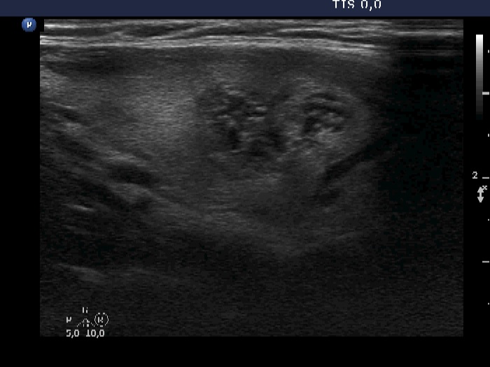
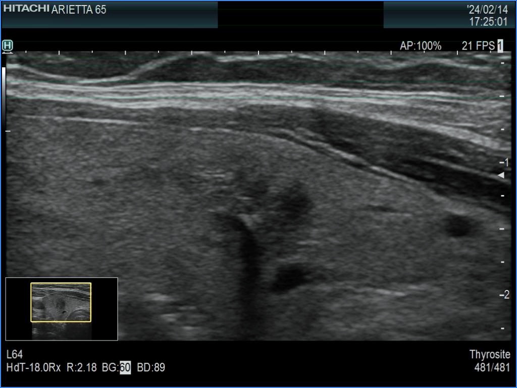
Second examination 4 years later (second row of images):
Clinical presentation: The woman came to routine follow-up. The thyrostatic therapy was stopped 3.5 years ago.
Palpation: The right lobe was suspicious having a not firm nodule.
Laboratory tests: TSH 0.29 mIU/L, FT4 16.2 pM/L.
Ultrasonography. The right lobe had multiple cystic nodules next to each other. The nodules displayed echogenic figures, which were caused by posterior enhancement. The presence of multiple nodules next to each other made the appearance lobulated. The dimensions of the nodular mass were 15x12x19 mm, width, depth, length, respectively.
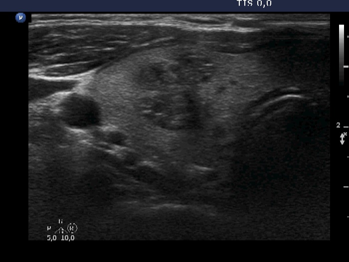
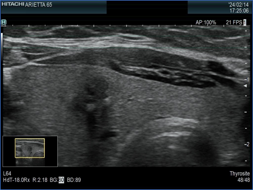
Ten years after the first examination (third row of images):
Clinical presentation: The patient came to a follow-up. She had no complaints.
Palpation: no abnormality.
Laboratory tests: TSH 1.38 mIU/L.
Ultrasonography. The nodule has significantly decreased in size, the dimensions were 7x7x6 mm, width, depth, length, respectively. The borders became irregular, lobulated while the echogenicity became hypoechoic.
Suggestion: ultrasound in 5 years.
Comment.
-
If we would meet the patient for the first time at the time of the last visit, the ultrasound presentation would cause concern. The nodule has lobulated margins, this is moderately or deeply hypoechoic. Considering the previous examinations, the lobulation is caused simply by the shrinkage of the nodule. So, this should not be considered as pathological lobulation.
-
I am not able to give an exact explanation for the change of echogenicity. Nevertheless, this not infrequently happens in nodule which decrease in size over time.
-
The appearance of macrocalcification is the sign of the ongoing degenerative process.