
|
|
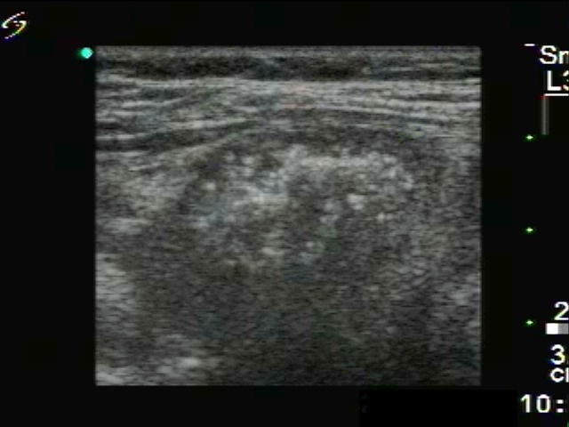
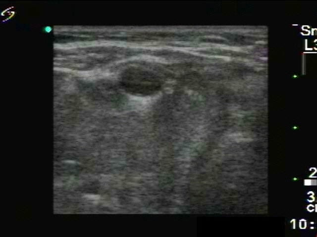
Intranodular hyperechogenic figures - case 1774
|
|


Clinical presentation: A 40-year-old woman was referred for evaluation of a nodule discovered 3 months earlier. The patient told us that the nodule has grown since its discovery.
Palpation: Hard nodules were palpable in both thyroids, enlarged lymph nodes were also present on the right side of the neck.
Functional state: hypothyroidism with TSH-level 13.0 mIU/L, FT4 10.1 pM/L.
Ultrasonography: There were multiple hypoechogenic nodules in both lobes. The lesions had hyperechogenic granules within an echonormal background.
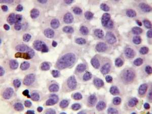
Cytological picture. There is no colloid in the background. Naked enlarged nuclei occur in solid nests and are dissociated without any structure. Thyrocytes are relatively small, elongated. The chromatin structure is irregular. A few enlarged cells, typical triangular forms with eccentric nuclei can also be found.
Cytological diagnosis: suspicion of malignancy. Taken the US pattern into account, it is medullary cancer with great probability.
Histopathology disclosed medullary cancer.
Comment. The hyperechogenic granules seen on ultrasonography correspond to amyloid deposits.