
|
|
Halo sign and vascular pattern of nodules - case 2248
|
|

Clinical data: A 77-year-old woman was referred for evaluation of a thyroid nodule discovered on screening.
Palpation: a moderately firm nodule in the upper part of the left lobe.
Laboratory test: TSH 1.08 mIU/L.
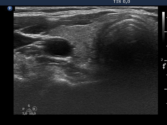
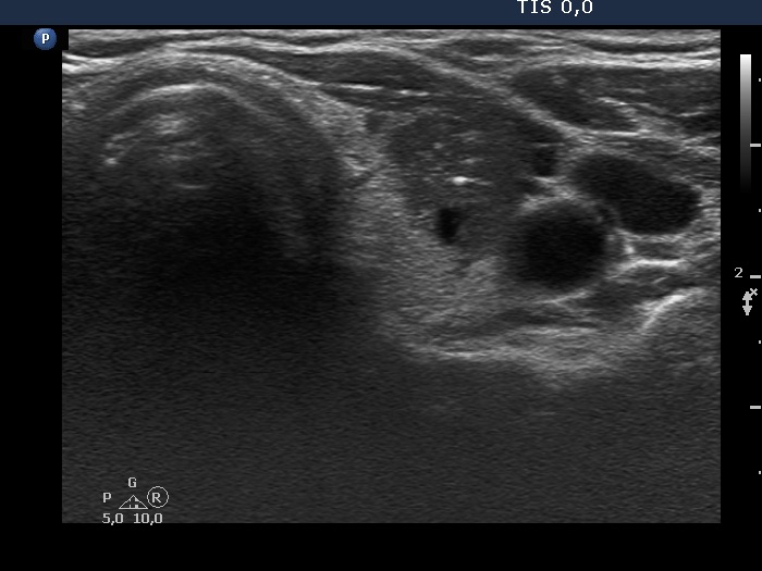
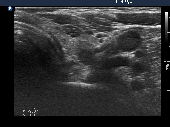
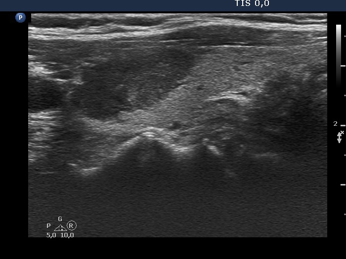
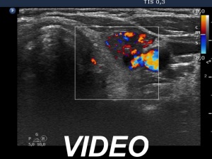
Ultrasonography. The thyroid was echonormal. There were several nodules in the left lobe. The largest in the upper pole presented all but one of the possible suspicious characteristics, it was deeply hypoechoic, had irregular margins, a microcalcification and showed abutting and bulging contours. Moreover, the intranodular vascularization was irregularly increased. One of the other lesions presented back wall figures while the third one showed taller-than-wide shape.
Cytology performed from the deeply hypoechoic nodule and resulted in benign lesion.
Suggestion: ultrasound and repeat cytology in a year.
Comment. The negative cytology in such ultrasound presentation does not fully exclude malignancy.