
|
|
Parathyroid lesions - case 1269
|
|


First examination (1st row of images)
Clinical presentation. A 18-year-old woman requested a screening because her mother was operated on a multinodular goiter. She had no complaints.
Palpation: no abnormality.
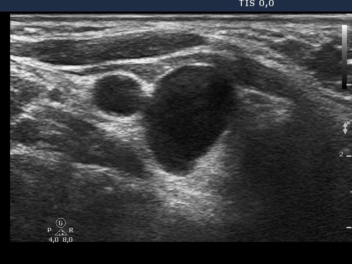
Ultrasonography. The thyroid was echonormal. There was a cystic lesion in the lower-dorsal part of the right lobe.
3 mL watery fluid was aspirated. There were no cells on the smear. The parathormone level of the cystic fluid was 495 pg/mL.
Functional state: Both the thyroid and the parathyroid functions were normal - TSH 1.06 mIU/L, parathormone level 22 pg/mL (normal value: 15-65).
Combined cytological-clinical-sonographic diagnosis: parathyroid cyst.
Second examination 2 years later (2nd row of images)
Clinical presentation. The patient had no complaints.
Palpation: no abnormality.
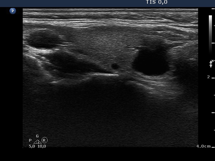
Ultrasonography. The pattern was essentially the same. The cyst has recurred.
1.8 mL watery fluid was aspirated.
Suggestion: ultrasound examination every second year.
Comment. The watery color of the cystic fluid is very specific for parathyroid origin.