
|
|
Thyroid cancers - case 1510
|
|


Clinical presentation: A 61-year-old woman referred for an evaluation of a rapidly enlarging bilateral thyroid mass. The tumor had evolved over 2 months.
Palpation: Both lobes were very hard, not freely moveable.
Functional state: hypothyroidism (TSH-level 11.8 mIU/L, FT4 13.8 pM/L, aTPO > 1000 U/Ll).
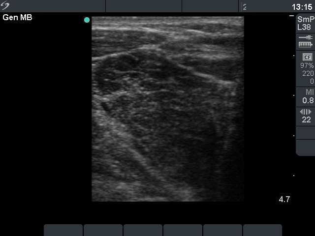
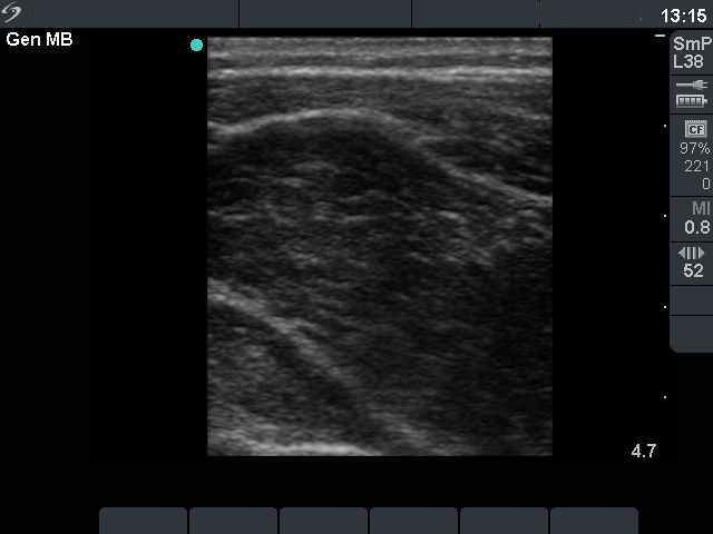
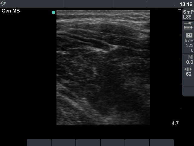
Ultrasonography. Both lobes were extremely enlarged, the largest diameter of the right lobe exceeded 12 cm. The whole thyroid was hypoechogenic and displayed fibrotic changes. No vascularization could be detected on Doppler mode.
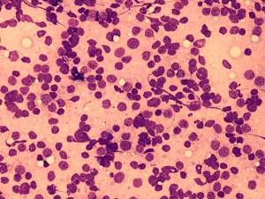
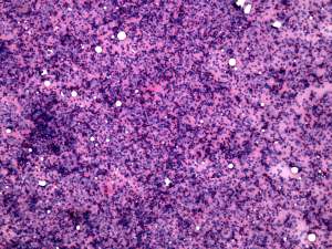
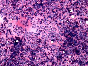
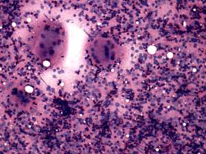
Cytological picture corresponded to Hashimoto's thyroiditis.
Final combined clinical-ultrasound-cytological diagnosis: suspicion of a MALT-type lymphoma.
Biopsy and histopathology: MALT-type lymphoma of the thyroid.
Comments.
-
The clinical presentation and patient history are the essence of the diagnosis of a MALT lymphoma of the thyroid. The role of cytology in such cases is the exclusion of an anaplastic carcinoma and to raise the suspicion of lymphoma.
-
The cytological pattern of Hashimoto's thyroiditis and MALT lymphoma is essentially the same. They differ in clinical appearance.