
|
|
Thyroid cancers - case 617
|
|


Clinical presentation: A 21-year-old woman was referred for an evaluation of hoarseness.
Palpation: no abnormality.
Functional state: euthyroidism (TSH-level 2.91 mIU/L).
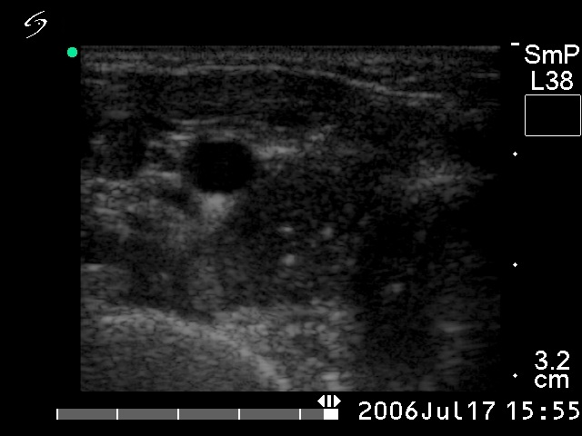
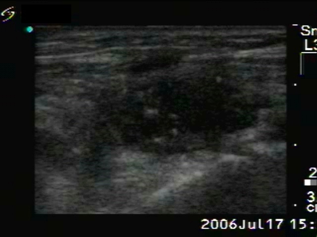
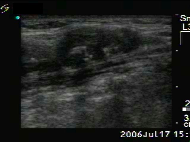
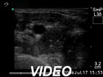
Ultrasonography. The right lobed was composed of a hypoechogenic nodule with hyperechogenic foci. The latter were greater than foci of a microcalcification and did not exhibit acoustic shadow. This is the typical appearance of an amyloid deposit. The left thyroid contained a similar tumor. There was a metastatic lymph node in the right side of the neck.
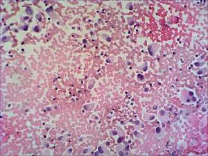
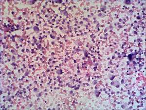
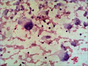
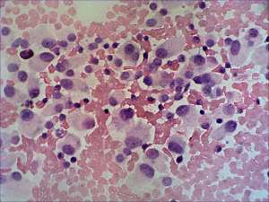
Cytological picture. There was no colloid in the background. There were enlarged atypical epithelial cells with eccentric nuclei without forming any structure resembling that of follicles. Many binucleated cells were found. Some nuclei contained intranuclear inclusion. Preliminary cytological diagnosis: suspicion of malignancy.
Calcitonin immunocytochemistry was positive and serum-calcitonin was elevated.
Final diagnosis medullary cancer.
Histopathology medullary cancer.
Comment. Naturally, the ultrasound images and video are technically out of date. Nevertheless, the specific ultrasound presentation of a medullary cancer is clearly seen.