|
|||||||||||||||||
|
Differential diagnostic problems: hyperthyoidism. Table 3
|
|||||||||||||||||
|
|||||||||||||||||
|
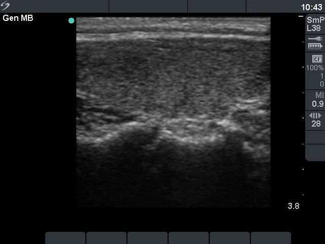
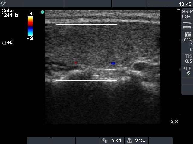
Compare the two series of pictures. The patient whose images are demonstrated on the left was treated with interferon and presented with a moderate degree of hyperthyroidism. Three important differences can be observed: the sonographic pattern is more hypoechogenic, the thyroid is more vascularized and larger in Graves' disease compared with Hashimoto's thyroiditis. |
|||||||||||||||||