
|
|
Chronic lymphocytic thyroiditis - Case 33.
|
|


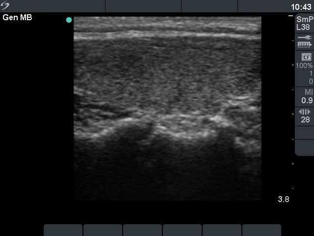
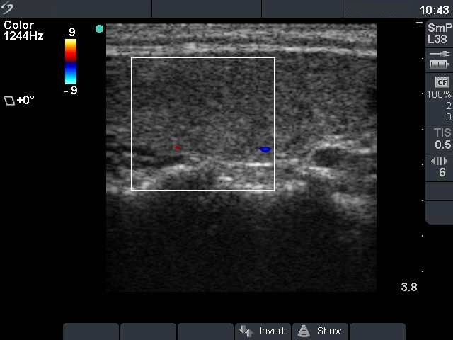


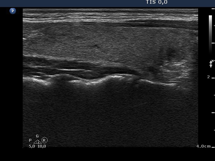
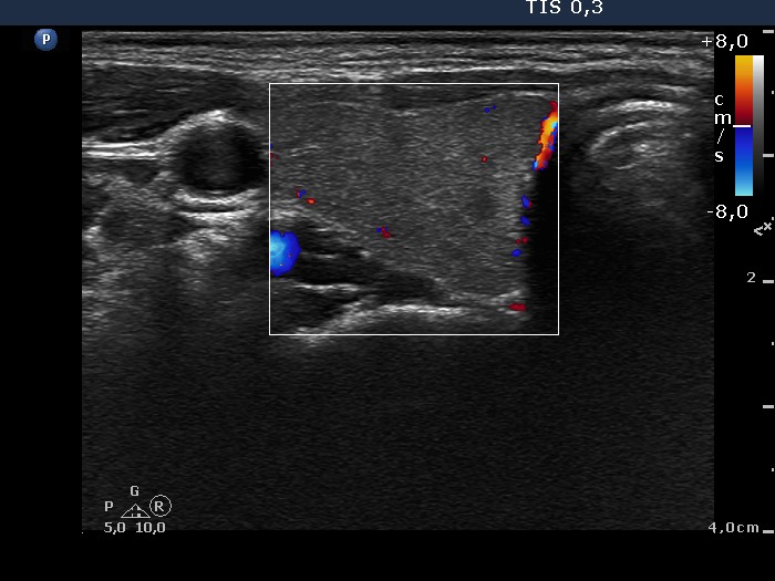

Follow-up investigation 2 years later (1st and 2nd rows of images)
Clinical presentation: a 37-year-old woman was treated with interferon because of her malignant melanoma. She had fatigue and palpitation in the previous 2 months.
Palpation: no abnormality.
Functional state: mild degree of hyperthyroidism (TSH 0.001 mIU/L, FT4 28.1 pM/L, aTPO 11 U/mL, TRAK 1.1 U/L (normal value below 1.5).
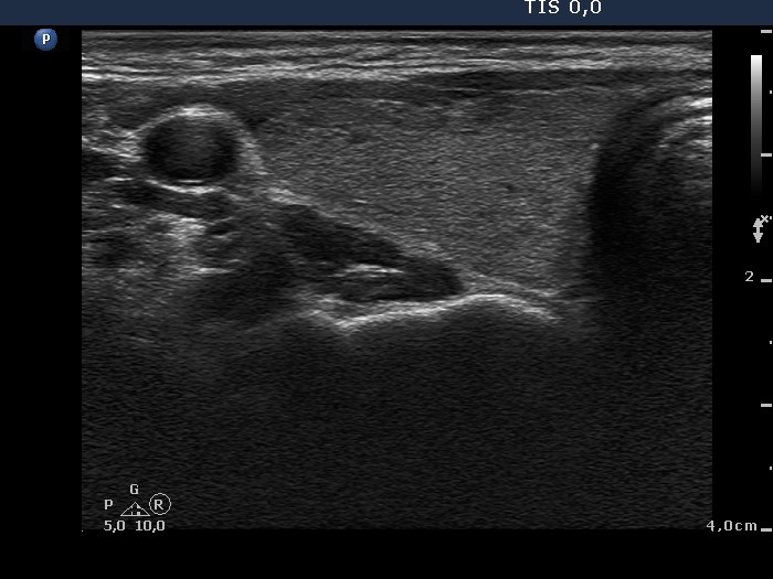
Ultrasonography: minimally hypoechogenic thyroids without any nodule. The vascularization was decreased.
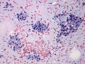
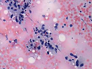
Cytology: resulted in lymphocytic thyroiditis.
Clinical diagnosis: hashitoxicosis.
We administered beta-blocking agent.
Three months later at the time of discontinuation of interferon therapy the biochemical status has not changed. On next follow-up investigation another 3 months later, the patient was clinically and biochemically euthyroid.
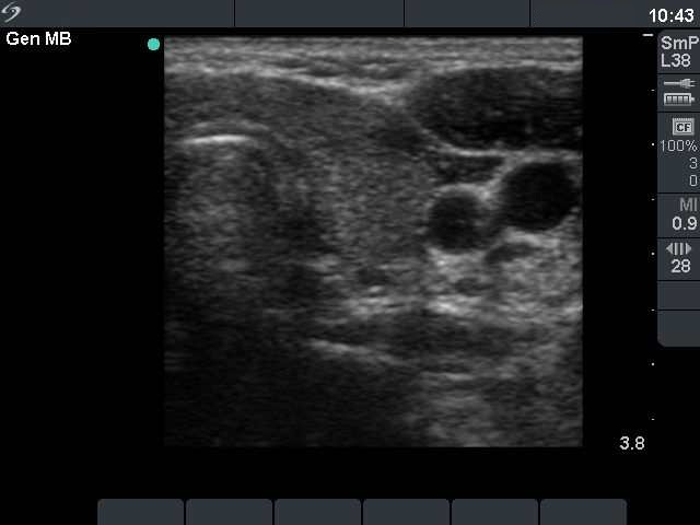
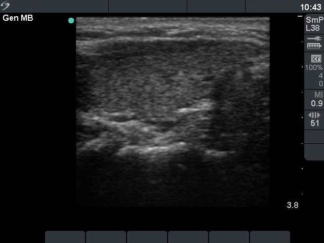
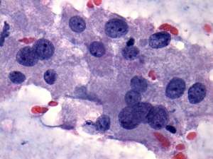
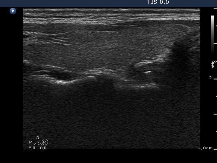
Follow-up investigation 2 years later (3rd row of images)
Clinical and biochemical data. The patient was well, clinically and biochemically euthyroid with TSH 2.81 mIU/L aTPO 5 U/mL)
Ultrasonography: the thyroids were practically intact.
Comment: we suppose that the patient had an underlying autoimmune thyroiditis despite her negative antibody results.