
|
|
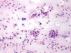
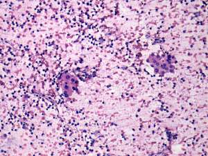
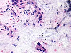
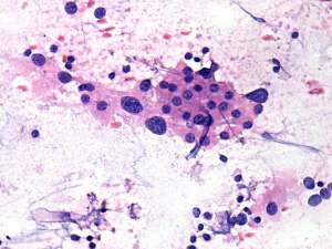
Chronic lymphocytic thyroiditis - Case 34.
|
|

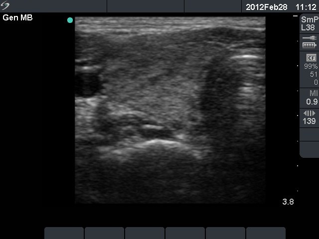
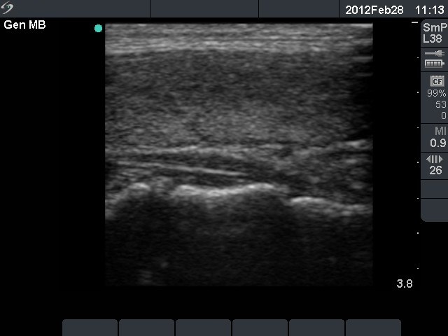

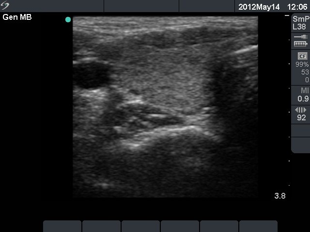


Initial investigation 5 months after delivery (1st and 2nd rows of images)
Clinical presentation: a 24-year-old woman was referred for an evaluation of weight loss. She had a delivery 5 months before the present examination.
Palpation: no abnormality.
Functional state: moderate degree of hypothyroidism with TSH 0.01 mIU/L, FT4 30.8 pM/L. TRAK 1.6 U/L (normal value below 1.5), aTPO 1 U/mL.
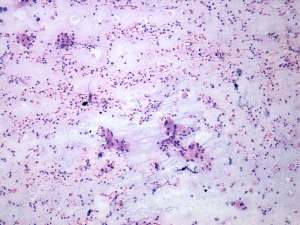
Ultrasonography: the thyroids were echonormal. The echogenicity index was around 15%. There was no nodule. The vascularization was average.Cytology resulted in Hashimoto's thyroiditis.
Clinical diagnosis: hyperthyroidism. Post partum thyroiditis.
We did not administer any therapy.
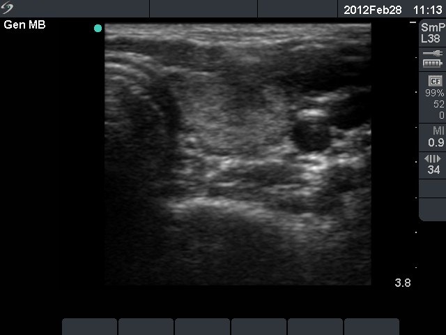
Follow-up investigation 2 years later (3rd row of images)
Clinical presentation: the complaints of the patient had decreased.
Palpation: no abnormality.
Functional state: euthyroidism with TSH-level 0.63 mIU/L, FT4 14.9 pM/L.
Ultrasonography: the thyroid became smaller. The echo pattern had not been changed on gray scale mode, but the vascularization had decreased.
We suggested follow-up examinations with yearly TSH determination in case of pregnancy.
Comment. The usual period of post partum hyperthyroidism is 5-10 weeks after delivery. Therefore in this patient the possibility of Graves' disease had to be considered. In such cases the FNAC may be of great help. This happened in this patient, cytology decided the differential diagnostic problem: the elevated FT4-level was caused by destruction and not by active hormone-producing disease.