|
|||||||||
|
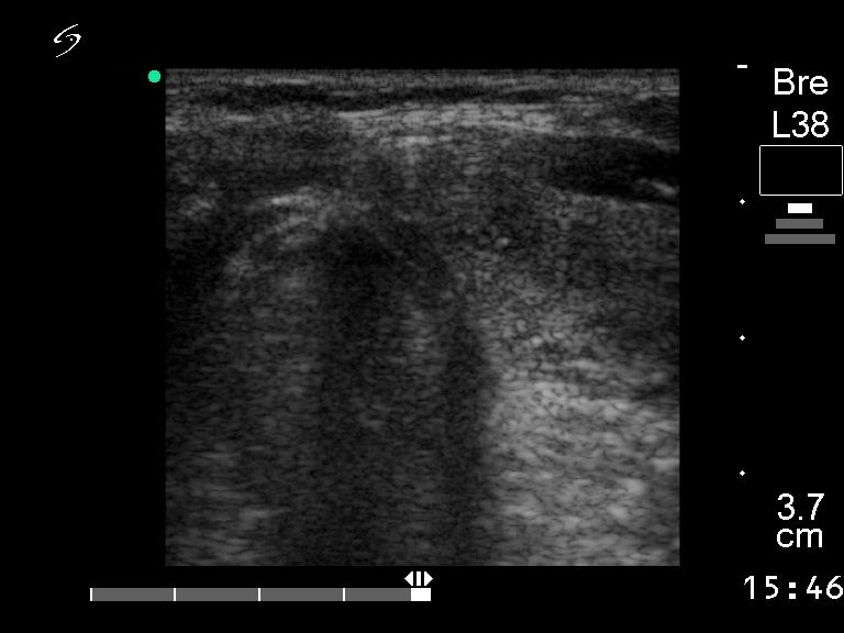
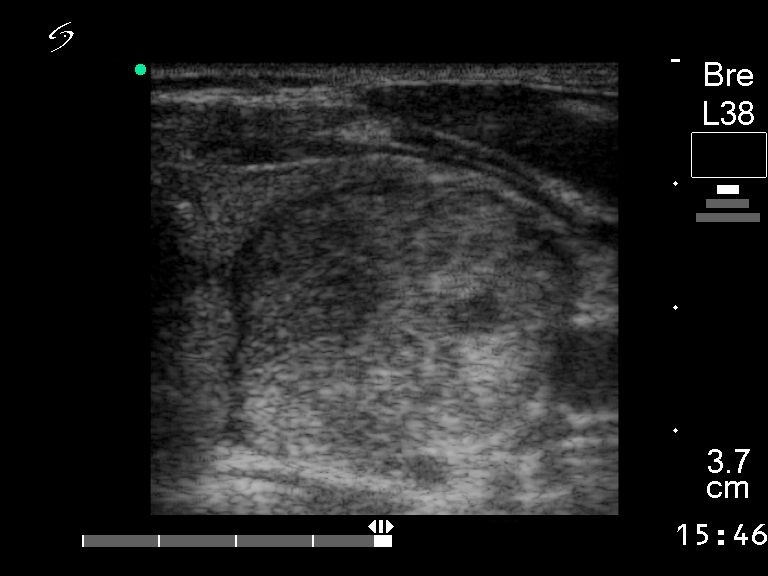
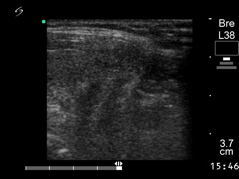
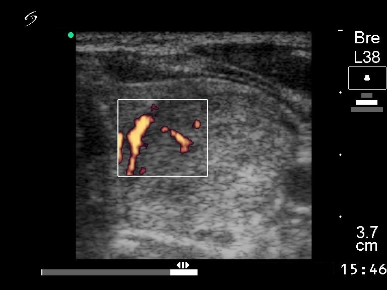
Hyperechogenic lesions in lymphocytic thyroiditis. Nodule or not nodule? Table 9 - Case 14 of oxyphilic adenomas
|
|||||||||
|
|||||||||
|
The images of the right lobe show the typical sonographic pattern of Hashimoto's thyroiditis: hypoechogenic thyroid with fibrotic changes. There was a hyperechogenic nodule within echonormal background in the left lobe.. Histopathology disclosed Hürthle-cell adenoma. Compare the size of this nodule with that of the lobule presented in the previous table. |
|||||||||