|
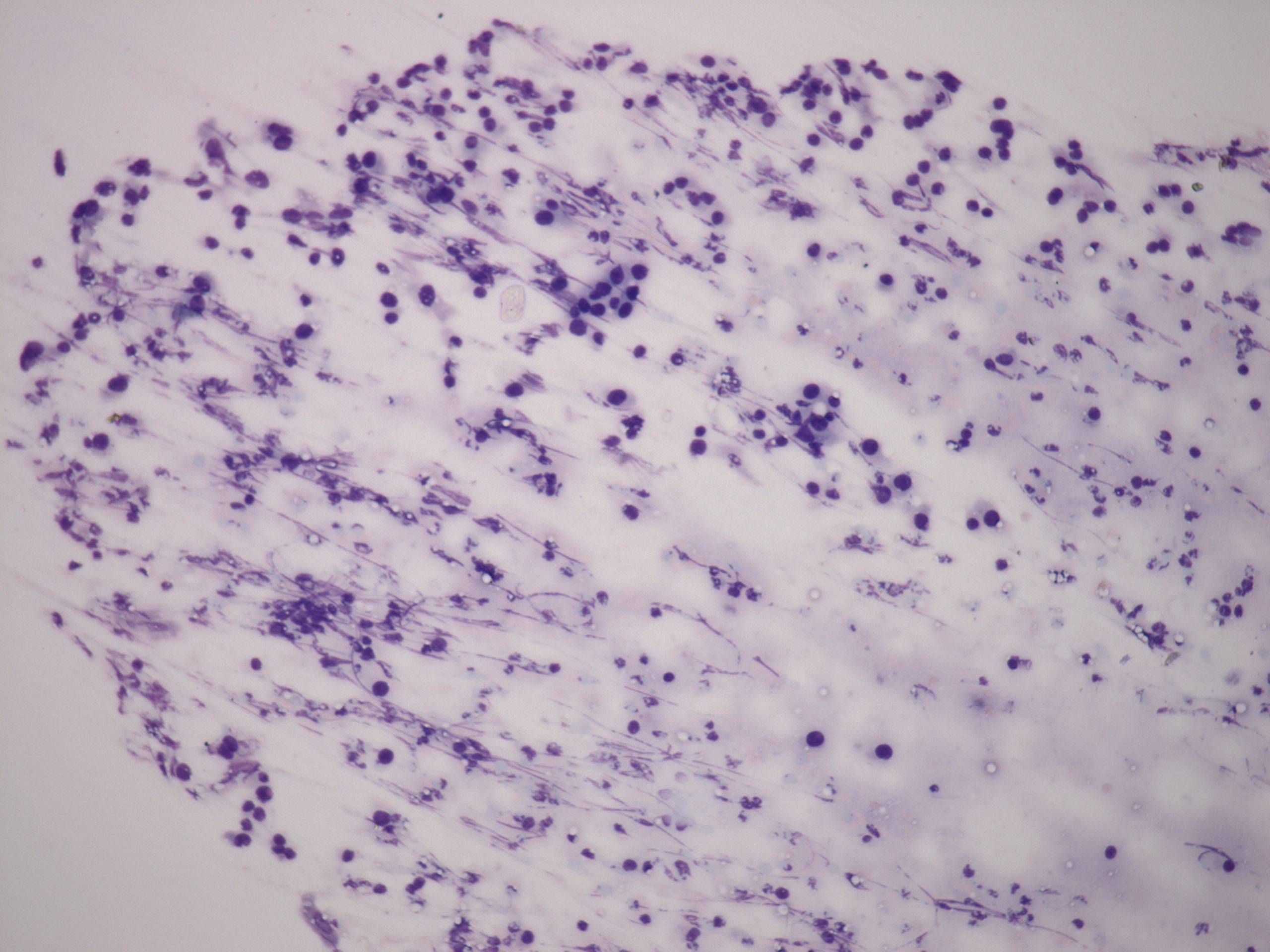
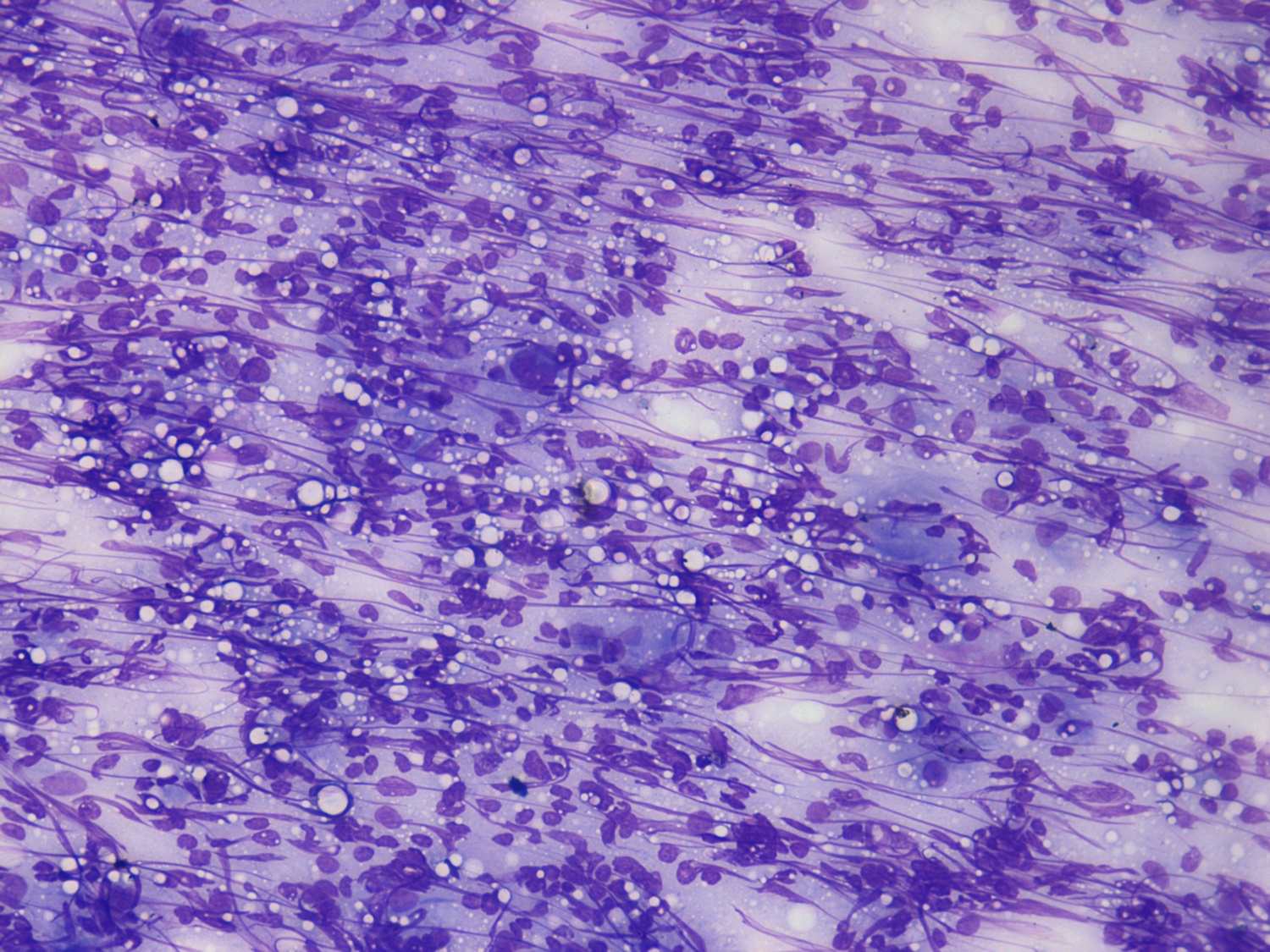
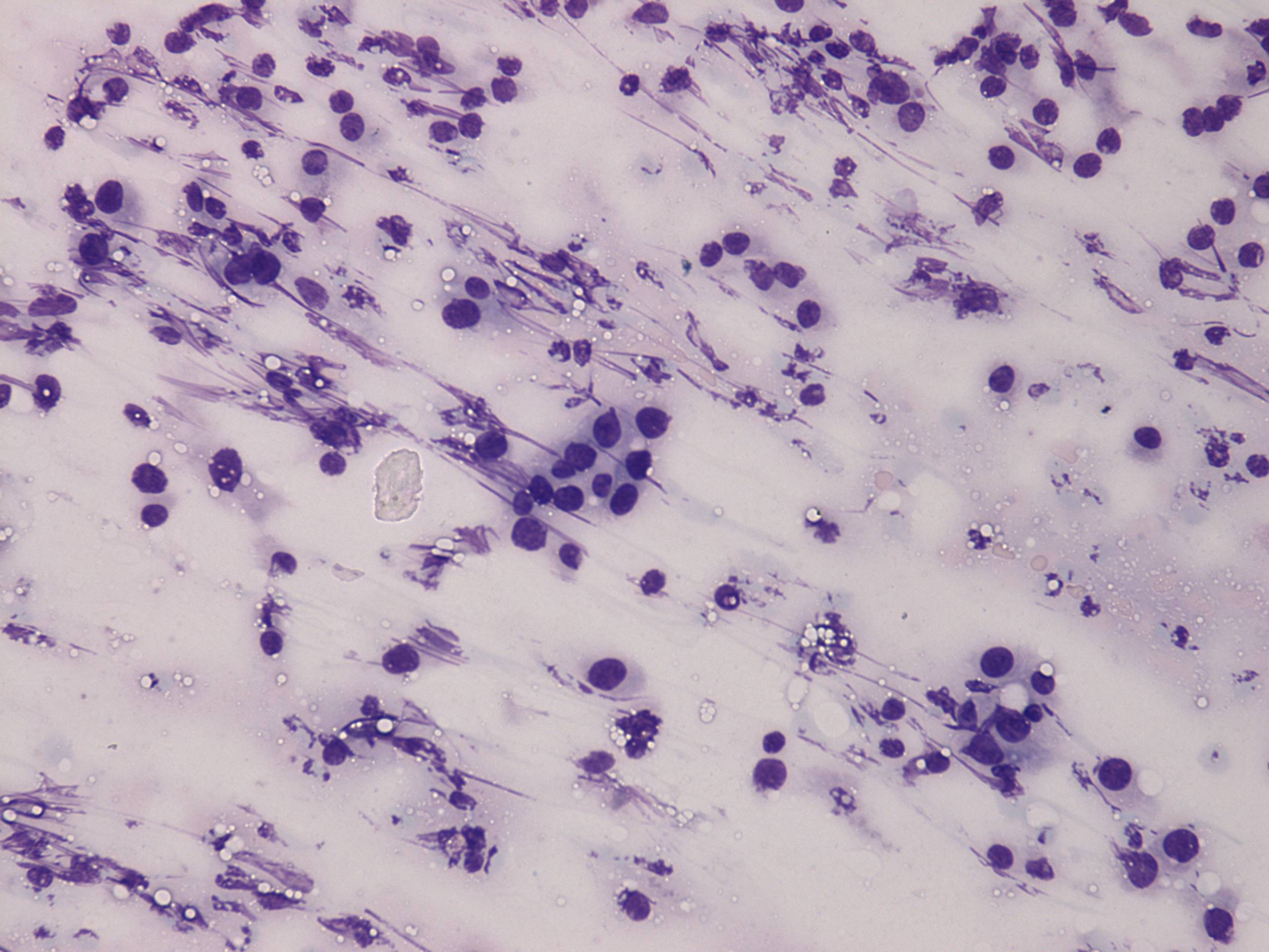
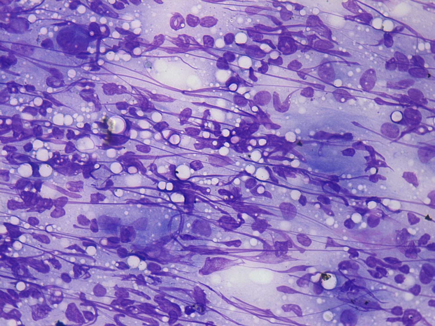
These two cases have a very similar cytological presentation. If we are
aware about the clinical picture, we can find that the 3 multinucleated
cells are partly composed of epitheloid cells in the left case, and we
will not misinterpret the epitheloid granuloma-like figure in the right
case. However, if the cytopathologist is not aware of the patient's
history, it is very difficult or even impossible to interpret the
cytological patterns correctly.
|